Wednesday, December 29, 2021

Gene editing 293T cell line facilitate cellular and molecular level researches!


Background

293T is a popular cell line for proof of concept. It can express SV40 T-antigen and contain SV40 origin of replication and promoter region. Many eukaryotic expression plasmids containing SV40 virus origin of replication, such as pcDNA3.1, can replicate in 293T cell line, so 293T cell line is widely used in virus packaging. 293T cell line also can be used for studying the expression of exogenous genes. Firstly, 293T cell line is easy to be transfected, and the transfection efficiency can be as high as 50% with calcium phosphate; Secondly, the protein expression level of 293T is high. The expressed protein can be easily detected by alkaline phosphatase analysis in 2-3 days after transfection. In addition, the transfection of the 293T cell line is one of the convenient ways to overexpress proteins and obtain intracellular and extracellular proteins. Thus, the 293T cell line has provided great help for a large number of cells and molecular level-related research. If you need to purchase 293T cells which have low passages, high activity and good cell condition, please click here >>


Detailed applications

293T cell line is widely used for researches, such as Lentivirus packaging and titer test, gene expression research, protein expression research, signaling pathway research, antibody screening, antibiotic screening, conditioned culture medium production, etc. Thus, 293T cell line is considered basic cell line by the researchers.


Applications of CRISPR/Cas9 technology in 293T cell line

CRISPR/Cas9 technology, as a popular gene-editing technology at present, has been well applied in 293T cell line. The stable cell lines are constructed through accurate gene-editing, which can be used to study apoptosis, functional genomics, signaling pathways, drug R&D, drug resistance mechanisms, drug response, and cell therapy. Ubigene’s developed CRISPR-U™ technology is 10-20 times more efficient than traditional CRISPR/Cas9 technology, and can greatly improve the recombination efficiency, which provides a powerful tool for gene-editing in 293T cell line.

So far, Ubigene has successfully generated a large number of gene KO 293T cell lines, the in-stock KO cell line is only 1880 USD. And we plan to construct ~900 genes knockout cell lines on the 293T cell line. These genes come from 8 popular signaling pathways and have high research value. If you are interested in our in-stock KO cell lines, please feel free to contact us!


Specific cases of gene-editing in 293T cell line

1. 293T cell line plays a key role in studying the effect of PLAC8 on the proliferation and migration of kidney cancer cells

The oncogene placental specific 8 (PLAC8) plays an important role in cellular processes and human diseases, but there are few studies on how PLAC8 affects the proliferation and migration of Human kidney cancer cells (KC). Therefore, Qin et al. selected 293T cell line as the target cell line to study the effect of PLAC8 on the proliferation and migration of 293T cell line. Firstly, they established two PLAC8 KO 293T clones with CRISPR/Cas9. In order to classify the characteristics of PLAC8 in the process of 293T cell line proliferation and migration, they conducted cell phenotypic tests such as cell counting, colony formation assay(cell proliferation), cell cycle, cell scratch test, and cell migration/Invasion(Ubigene provides various Cell Phenotypic Services). The molecular mechanism was studied by Western blot. The results showed that KO PLAC8 could inhibit the proliferation of the 293T cell line. In addition, the inhibitory effect of PLAC8 KO on 293T cell line proliferation was related to G2/M in the cell cycle. At the same time, PLAC8 KO significantly inhibited cyclin B1 and increased cyclin a. This shows that PLAC8 plays an important role in 293T cell line proliferation and migration, and provides an important research basis for further study of the effect of PLAC8 on human KC cells [2].


2. 293T cell line helps to build a quantitative model of recombinant protein production

293T cell line can express foreign genes. So it has been considered a powerful tool to produce and express specific protein products, and has been widely used to produce a variety of recombinant proteins. However, the recombinant protein yield of constructed cell lines is often inconsistent and difficult to quantify. Lo et al. used CRISPR/Cas9 to bind human ribosomal protein L13A(RPL13A) KI to a specific site (Fig. 3), so that transgene expression is driven by endogenous promoters to ensure consistent and predictable expression of recombinant proteins. The expression level can also be determined in advance by selecting promoters from genes with the required expression level. In order to quantify the expression of recombinant protein, protein quantitative reporter gene (PQR) is integrated between endogenous gene and exogenous gene. PQR equimolar produces endogenous protein, recombinant protein and fluorescence reporter gene, so the expression of target protein can be quantified by cell fluorescence intensity.[3] 

Ubigene specialized in gene KI cell line generation, as low as 9480 USD, it will come with a $2000 coupon for cell phenotypic services. For more details, please inquire >>


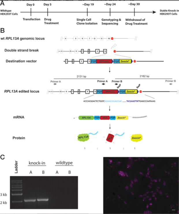
Figure 1 RPL13A KI 293T cell line generation


3. Study of telomerase activity inhibition by 293T cell line

All continuously proliferating cell lines, such as stem cells and cancer cells, have a mechanism to compensate for telomere wear during continuous division. In most cases, this requirement is met by telomerase. Somatic cell line cannot divide indefinitely due to the lack of telomerase activity, but the reactivation of TERT transcription in cell lines will enable them to continue to divide, which is a key step in the process of tumorigenesis. Therefore, the study of TERT expression is of great significance to understand the regulatory mechanism of telomerase activity under physiological and pathological conditions. Two point mutations in the promoter region of the human TERT gene (C-124T and C-146T) are highly recurrent in various cancer types and are associated with higher telomerase levels. Xi et al. modified the endogenous TERT gene with CRISPR/Cas9 and labeled the endogenous TERT protein or protein with localization tags. They generated a 293T cell line expressing FLAG-SNAP labeled TERT protein, which realized the effective immunopurification (IP) and subcellular localization of endogenous TERT. The results showed that telomerase was localized to only a few telomeres at any given time. They also generated HEK 293T and SCaBER cell line with a modified TERT promoter, suggesting that removing the C-124T mutation is sufficient to reduce telomerase levels and shorten telomeres. This method not only provides a useful tool for the study of telomerase biology, but also provides a general method to purify and visualize low abundance proteins and modify single base pairs at genomic sites with low editing efficiency.[4]


Based on over 5000 successful gene-editing cases, Ubigene provides high-quality gene-editing cell line services, including knockoutknockin, and point mutation.  We also offer stable cell line generation service for customers. If you are interested in one of our services, please feel free to contact us!


Tuesday, December 28, 2021

[Research highlight] Study the FOSL2's involvement in the regulation of muscle glycogen(MG) content, Ubigene can help!


Muscle glycogen (MG) is an important intermediate in the process of glucose metabolism, which can maintain the stability of blood glucose and regulate the balance of glucose metabolism. MG not only participates in maintaining the normal physiological activities of animals, but also affects the homeostasis, growth performance and meat quality of livestock and poultry after slaughter. However, the key genes regulating MG metabolism in poultry have not been determined.

Recently, Xiaojing Liu,etc. (Chinese Academy of Agricultural Sciences Pasturage Institute) published "FOSL2 Is Involved in the Regulation of Glycogen Content in Chicken Breast Muscle Tissue" in Frontiers in Physiology (2020 IF4.566), which confirmed the key role of FOSL2 in regulating glycogen content in chicken breast muscle tissue. This discovery is helpful to understand the metabolic regulation mechanism of muscle glycogen in chickens, and provides a new idea for the production of high-quality broilers and the formulation of comprehensive nutritional control strategies.


In previous studies, researchers found that FOSL2 is a candidate gene related to chicken muscle glycogen (MG) content, but the role of FOSL2 in regulating MG content is still unclear. Differential gene (DEGs) expression analysis and weighted gene coexpression network analysis (WGCNA) were carried out in the breast muscle samples of yellow chickens in high MG group (HMG) and low MG group (LMG). It was found that except FOSL2, there were some genes related to MG metabolic pathway, such as PRKAG3, CEBPBFOXO1, AMPK and PIK3CB; In addition, it was also found that FOSL2CEBPBMAP3K14 and other classical glycogen metabolism related genes were related to MG content in the same co-expression module by the WGCNA. The expression of FOSL2 was negatively correlated with MG content, and there was a possibility of interaction between FOSL2 and CEBPB.


The researchers further studied the effect of FOSL2 lentivirus overexpression (constructed by Ubigene) on glycogen content in vitro. The results showed that FOSL2 reduced the glycogen content in the chicken fibroblast DF1 cell line, which was consistent with the analysis results of transcriptome and WGCNA data. Ubigene provides plasmids construction and Virus Packaging services for customers globally, as low as $559.


The research results show that the key role of FOSL2 in regulating MG content in chicken cell lines. FOSL2 may negatively regulate MG content by regulating its downstream gene CEBPB, which is not affected by variety and gender. And it provides new understanding for the study of MG metabolic regulation.

Consequently, the main idea of this article is to find the candidate gene first, and then overexpress the target gene to study the regulation mechanism and gene function. Obviously, different idea of research needs to modify candidate genes in different ways. 


Ubigene provides gene-editing services for customers all over the world. Ubigene not only provides plasmids construction and Virus Packaging services, but also offers custom stable cell line generation services to meet the different research needs of customers! For information, please inquire >>


Reference 
Liu X, Liu L, Wang J, Cui H, Zhao G, Wen J. FOSL2 Is Involved in the Regulation of Glycogen Content in Chicken Breast Muscle Tissue. Front Physiol. 2021 Jul 6;12:682441. doi: 10.3389/fphys.2021.682441. PMID: 34295261; PMCID: PMC8290175.

Monday, December 27, 2021

How does CRISPR/Cas9 work for RKO cell line research? |Ubigene


RKO cell line is a low differentiated colon cancer cell line developed by Michael Brattain. It contains wild-type P53, but it lacks human thyroid receptor nuclear receptor (h-TRβ1). RKO cell line is the parental cell line of RKO-E6 and RKO-AS45-1, and its P53 protein level is higher than that of RKO-E6 cell line. This cell line can form tumor in nude mice or colony in soft agar. RKO cell line is widely used in molecular, cell biology and biomedical basic research, such as proteomics, genomics, cell line research, DNA, RNA and genetics. It is also used in popular biomedical industry, such as antibiotic screening, antibiotic metabolism and toxicology research, cancer drug research, etc. Ubigene provides wild-type RKO cell line with good condition, and can be applied to various gene-editing experiments, which is convenient for constructing various RKO cell line models, such as Knockout, Knockin, and point mutation or other stable cell lines


  Detailed applications of RKO

For cancer research, the results obtained from cell line research can be validated by RKO cell line;

Predict the response and rejection of individual samples to select the most effective anticancer drugs;

Screening the most selective or toxic new drug compounds;

As a recognition target for antitumor antibiotic screening and model construction.

  Applications of CRISPR/Cas9 technology in RKO cell line

CRISPR/Cas9 system is changing many biomedical research prospects, including cancer research. By flexibly inducing DNA double-strand breaks, cancer mutations can be transfected into cell lines in vitro or in vivo. However, not all mutations have the same effect on tumorigenesis. It is troublesome to distinguish between basic mutations and bioinert mutations in tumor growth and survival. Sayed et al. proposed a method to screen the functional correlation of high-throughput mutations in RKO cell line. They used CRISPR/Cas9 system to detect cancer vulnerability in colorectal cancer cell lines, and used positive or negative sieves to isolate cell lines with specific phenotypic changes from the whole collection population to try to identify new cancer driven mutations. They designed 100 high-quality sgRNAs that can specifically cleave mutations in RKO cell line. A multifunctional lentivirus library containing these sgRNAs was then generated and used for joint screening to detect the possible growth conditions of these mutations. Genomic DNA was collected at different time points, sgRNA was amplified and purified by PCR, and sgRNA was counted quantitatively by deep sequencing. The results showed that in RKO cell line, two sgRNAs targeting the same mutation (UTP14A:S99delS) would be depleted over time (Fig. 1A). The experiment results confirmed that the inactivation of this mutation impaired cell growth, so Sayed et al. considered UTP14A:S99delS as a driving mutation in RKO cell line (Fig. 1B). The above research results show that CRISPR/Cas9 system can realize large-scale functional separation of cancer mutations, which obviously provides effective help for future cancer research and treatment [2]Ubigene has developed the exclusive CRISPR-U™based on CRISPR/Cas9 technology, CRISPR-U™technology has more efficient gene cutting efficiency and can improve gene-editing efficiency by at least 10 times. Ubigene provides CRISPR-U™ gene-editing service all over the world, including KO, KI, and point mutation, plasmids construction, custom stable cell line generation, etc.


A                              B

Figure 1

  Specific cases of gene-editing in RKO cell line

DIP2C deletion plays an important role in RKO cell line in cancer research

Disco-Interacting Protein 2 Homolog C(DIP2C), a non characteristic gene highly expressed in human tissues and adult tumors, was identified as a putative cancer gene in hormone mutation analysis within the exome. In order to study the role of DIP2C deletion in human cancer and determine the process affected by the activity of this gene, Larsson et al. constructed a human DIP2C KO RKO cell line with CRISPR/Cas9 technology. The results showed that the knockout of DIP2C in RKO cell line resulted in cell enlargement and growth retardation. Larsson et al. revealed 780 genes that were affected by DIP2C deletion, including the tumor suppressor genes encoding CDKN2A, ZEB1 encoding epithelial mesenchymal transition (EMT) regulatory factors, and CD44 and CD24 encoding breast cancer stem cell lines. DNA methylation analysis showed that more than 30000 gene loci were affected by differential methylation, most of which were hypomethylated after DIP2C deletion. The changes of DNA methylation in promoter region are closely related to the changes of gene expression. RKO cell line after DIP2C knockout have higher wound closure capacity, so the deletion of DIP2C will trigger a large number of DNA methylation and gene expression changes, cell line aging and epithelial mesenchymal transformation in cancer cell lines [1].


Figure 2 DIP2C KO leads to the change of gene expression


Screen the most resistant factors and promote the R & D process of inhibitors

Necrotic cell death pathway is a key component of human pathogen defense. It can be abnormally inhibited in the process of tissue homeostasis, resulting in many types of tissue damage and diseases. Although the formation mechanism of necrotizing body kinase signal complex containing RIPK1、RIPK3 and MLKL has been well known, other mechanisms of its regulation and effector function remain to be found. Callow et al. conducted whole gene knockout through CRISPR/Cas9, screened 19883 mouse protein coding genes, screened out factors resistant to cell necrosis through sgRNA reference library obtained by large-scale transduction of RKO cell line, and then identified 112 necrosis regulatory factors and mediators, including 59 new candidate pathway components. It was found that cell growth was not affected without necrosis induction. They further suggest that some tissue-specific or stress response pathways may prevent regulated cell death by regulating RIPK1. The above research results show that the further exploration of RIPK1 function plays an important role in promoting the development of new therapeutic intervention strategies or clinical inhibitors [3].


Figure 3 Antibiotic resistance factor screening


Whole genome knockout RKO cell line provide crucial help for radiotherapy CRC

Colorectal cancer (CRC) ranks among the most frequently diagnosed cancer and is one of the leading causes of cancer-related death worldwide. In addition to surgical resection and chemotherapy, adjuvants of radiotherapy also play a key role, which can prolong the survival rate of patients. However, in the course of radiotherapy, about 50% of patients will develop resistance to radiotherapy adjuvants, resulting in unsatisfactory curative effect, recurrence and metastasis. Therefore, identifying the potential mechanism of radiation resistance can improve the treatment outcome of patients with colorectal cancer. Yu et al. conducted genome-scale CRISPR knockout screening in RKO cell line and combined with NGS sequencing to explore the regulatory factors involved in radiation resistance of colorectal cancer, and finally obtained three candidate genes. They found that microRNA-5197-5p (miR-5197) could significantly enhance the cytotoxicity of IR to RKO. Through further mechanism research, Yu et al. proved that miR-5197 directly targets CDK6 and inhibits its expression in RKO cell line, which can induce cell cycle arrest in G1/S phase and inhibit cell division. Therefore, CDK6-5197 enhances radiosensitivity. The results show that miR-5197 may be a key factor in regulating the radiosensitivity of CRC cell line and help to develop treatment strategies for CRC patients resistant to IR [4].


Figure 4 Candidate genes related to CRC radiation resistance were identified by genome-wide CRISPR knockout screening


Ubigene has successfully modified gene from more than 100 cell lines with the developed CRISPR-U™  technology, including RKO cell line.  And Ubigene has successfully  generated over 5000 KO cell lines for the researchers all over the world. Ubigene is  now providing a time-limited promotional price for all gene-editing services.  If you want to get more information, please feel free to contact us!


Friday, December 24, 2021

purchasing cell lines, you have to know these 4 tips .


You must have searched “how to purchase xx cell line” online. So how to purchase a good cell line via various advertisements? Here are some useful tips for purchasing cells.


Tip 1: Clear cell information

If you want to purchase the cell lines, of course, you should know which cell line you want to buy, so it is the most common requirement to know the information of the purchased cell line. The name of the cell is the most important information. It often contains the information such as species and tissue source, so that we can quickly get the identity of cells. Then you need to know whether your target cell is a primary cell or a cell line. Primary cells can maintain their original genetic characteristics and are suitable for experimental research with high requirements such as antibiotic screening, cell differentiation and transformation; The cell lines have the advantages of low price, easy to culture, unlimited passages; These two kinds of cells are not only different in application, but also in price. Cells need to be selected according to your experimental needs. In this way, you can accurately express the purchase demand and avoid wrong cell purchasing and delivering due to the communication problems. Ubigene has two cell banks, wild-type cell line bank and stable cell line bank. The common information of cell line has been shown in our products webpage, and you can download the introduction for free to view the STR Authentication data of the cell lines, and avoid purchasing or recognizing the wrong cell lines.

Click here to select >


Tip 2: STR Authentication

Many academic journals require researchers to provide STR Authentication results of cell lines in recent years, including NIH, ATCC, Nature and Science.Many researchers use cross-contaminated or misidentified cell lines, resulting in unacceptable results, such as unrepeatable experimental results and negative research conclusions, which means they waste a lot of time for this small error. Especially, it will lead to the serious consequences in clinical cell therapy, just because of the wrong research conclusions.


Cell identification method: STR Authentication is one of the most effective and accurate methods for cell cross-contamination and characteristic identification. Many academic journals require researchers to provide STR Authentication results of cell lines recently. Therefore, it is very necessary to ask whether STR Authentication report is provided and check is the STR Authentication results are true. If the experiments of several months or even years is about to end without STR Authentication, it may lead to the negative conclusion, such as the waste of time and funds, and everything has to start from the beginning. All human and mouse cell lines  in Ubigenes wild-type cell line bank provide STR Authentication reports for free to ensure cell identity.


Tip 3: Strict QC

First, it is important to know whether cell lines are contaminated by bacteria, fungi and mycoplasma. After mycoplasma contamination, the expression of DNA, RNA and protein in cell lines will change. When the contamination is serious, the cell lines will grow slowly, cause the cell morphology change, and cause the cell pathological changes. Mycoplasma contamination is relatively difficult to deal with. So we should ensure cell lines are not contaminated by mycoplasma when we puchase the cell line.


Secondly, we need to pay attention to the passage level of cell lines. With the increase of passage, cell lines will have problems such as bad growth condition and mutation, unpredictable experiment results, such as changes in protein expression. Try to use low passage, healthy and active cell lines in order to ensure the reliability of experiment.


Ubigene regularly carries out strict microbial detection on all cell lines in our cell bank, and ensures low passage, good cell condition. It is even suitable for cell transfection and antibiotic screening during the experiments, such as gene-editing, stable cell line generation, etc.


Tip 4: Professional technical guidance

When you confirm the above 3 tips, you can ensure that the cell line you purchase is good. Even if unluckily, the cell line is not good from some companies, you can better protect your rights. On the contrary, some researchers purchase the cell lines in good condition, but they can't better culture the cells. They may think it's the problem of the cell lines. And some researchers ask for help about this problem. Some cell lines seem in good condition, but once doing gene-editing, the cell lines may be in bad condition after cell transfection or antibiotic screening due to lack of experience, cell lines are easy to die, the transfection rate is low, or the efficiency of gene-editing is low. Generally, some companies will provide some technical guidance, but it is just about the common culture of cell lines. For more gene-editing experiments, it may be more reliable to find a company specializing in gene-editing services.


Ubigene focuses on providing professional gene-editing services and products. We take the high standard of "suitable for all gene-editing experiments, and also provides free technical guidance for gene-editing! Make your gene-editing experiment easier!

Ubigene, make genome editing easier!

Thursday, December 23, 2021

ATP7B knockout cell line, a good helper on the treatment of WD |Ubigene

Hepatolenticular degeneration, also known as Wilson's disease (WD), is a rare autosomal recessive disorder of copper metabolism, and the pathogenic gene is ATP7B. The ATP7B gene, located on chromosome 13 (13Q14.3), encodes a 1411 amino acid Cu transporter P-type ATPASE. Mutation of ATP7B gene results in weakened or lost ATPASE function, resulting in decreased synthesis of ceruloplasmin (CP) in serum and impaired biliary duct Cu discharge, and deposition of Cu ions in liver, brain, kidney and cornea. It would causes progressive cirrhosis, extrapyramidal symptoms, psychiatric symptoms, kidney damage, and corneal pigmentation ring (Kayser-Fleischerring, K.F ring), resulting in Wilson's disease. The worldwide incidence of WD is 1/30 000 ~ 1/100 000, and the prevalence of WD is about 1/90. In Asia, the most common type is R778L, but the pathogenesis of this mutation remains unclear. This disease is more common in China, which is more common in young people and slightly more common in males than females. If not properly treated, WD can cause disability or even death. WD is also one of the few treatable neurogenetic diseases. The key is early detection, early diagnosis and early treatment.


The combination therapy of MT1X induction and Cu chelation provides an effective idea for the treatment of Wilson's disease.

Cu chelators and zinc salts are the two most important drugs used in the treatment of WD patients; However, the molecular mechanisms associated with ATP7B expression in these drugs have not been identified. A targeted knockout of ATP7B (KO) was established in the most widely used human hepatoma cell line, HepG2 for molecular studies of the pathogenesis and treatment of the disease. KO cells showed similar growth, Cu uptake, release, and gene expression as compared to parental cells. However, in the presence of Cu, morphological changes, oxidative stress, apoptosis, and loss of viability were observed. Induction of metallothionein (MT1X) after Cu exposure was significantly reduced in KO cells. Following zinc treatment, MT1X expression was strongly induced and a high percentage of KO cells could be rescued from Cu induced toxicity. D-penicillamine treatment had a minor effect on the viability of KO cells whereas the parental cell line showed a pronounced improvement. Combined treatment displayed a highly synergistic effect in KO cells. The data suggest that zinc has a previously unrecognized effect on the viability of hepatocytes that lack ATP7B due to a high induction of MT1X expression that compensates low gene expression after Cu exposure. A combination therapy that simultaneously targets at MT1X induction and Cu chelation improves the overall survival of hepatocytes for most efficient therapy of patients with WD. KO cells played a key role in this study, Ubigene now has nearly 2,000 types of KO cells in stock, covering thousands of genes from 8 popular signaling pathways, popular immune checkpoints and diseases (most are cancer disease).


Verify the effects of Cu on lipid homeostasis in intestinal cells with ATP7B KO cell line.

In another study, Guttmann et al. constructed ATP7B knockout cell lines in human intestinal cacO-2 cells to determine the relationship between Cu and lipid homeostasis and metabolism in intestinal cells. Caco-2 cells were transfected with plasmids containing endonuclease Cas9 and the gRNA target to exon 2 of human ATP7B. The results showed that KO cells had increased sensitivity to Cu, increased intracellular Cu storage, and induced genes regulating oxidative stress. The results showed that the sensitivity to Cu and intracellular Cu storage had increased, and induced genes regulating oxidative stress in KO cells. The chylomicron structural protein ApoB48 was significantly down-regulated by Cu in KO cells. Apolipoprotein ApoA1, ApoC3, and ApoE were induced by knockout of ATP7B. Cu enhances the formation of small lipid droplets and decreases the formation of large lipid droplets. At the same time, Cu decreased the storage and secretion of triglycerides (TG). The KO cells exposing in oleic acid (OA) leads to enhanced TG storage. In a word, The results suggest that Cu inhibits TG adipogenesis in the intestine, while ATP7B deficiency leads to TG storage induced by OA.


The mouse model of ATP7B gene R778L and P992L point mutation was constructed by CRISPR/Cas9 technique.

Dong et al. used CRISPR/Cas9 technology to target Arg778Leu and Pro992Leu, which are the common mutation sites of ATP7B gene in WD patients in China, and injected Cas9 mRNA into mouse single-cell embryos by microinjection. Finally, a mouse model simulating ATP7B gene R778L and P992L point mutations was successfully constructed. The clinical phenotypes of the two mutant mice were observed from the physiological and pathological perspectives, and their phenotypes were verified to conform to the clinical symptoms of WD patients. It was found that the serum ceruloplasmin level of R778L point mutant mice was significantly lower than that of wild-type mice, while the serum non-ceruloplasmin binding Cu content of R778L and P992L mice was significantly higher than that of wild-type mice. At the same time, these mice showed pathological liver damage, while R778L mice showed behavioral abnormalities. This study accurately constructed a point mutation mouse model of WD in Chinese population, providing an ideal tool for the pathogenesis and gene therapy research of WD.


"Make gene editing easier!" has always been Ubigene aspiration. Ubigene developed the CRISPR-U™ technology, which is greatly improve the efficiency of homologous recombination. With CRISPR-U™ , Ubigene has successfully generated over 5000 KO cell lines for the researchers all over the worldIn addition, Ubigene now has nearly 2,000 types of KO cells in stock. For more details, please feel free to contact us!


Reference:

[1]ursimran C , Nadine S , Vanessa S , et al. The Effect of Zinc and D-Penicillamine in a Stable Human Hepatoma ATP7B Knockout Cell Line[J]. Plos One, 2014, 9(6):e98809.

[2]Guttmann S , Nadzemova O , I Grünewald, et al. ATP7B knockout disturbs copper and lipid metabolism in Caco-2 cells[J]. PLoS ONE, 2020, 15(3):e0230025.

[3]Jianjian D. Construction of Wilson's disease mouse model using CRISPR/Cas9 technique and study on gene therapy[D].University of Science and Technology of China, 2021.DOI:10.27517/d.cnki.gzkju.2021.000522.

Tuesday, December 21, 2021

Optimized culture medium,boost single-cell clone formation!


What's the difficulty of cell culturing


Cell culturing plays an important role in gene-editing experiment. By simulating the internal environment (asepsis, suitable temperature, pH and certain nutrition, etc.) in vitro, cells can survive, grow, proliferate and keep their main structure and function. It seems simple, but this process is a real headache for many researchers. For example, adherent cells do not adhere, suspended cells become clustered, even inadvertently, cells may die...


Some experiments have higher requirements on the cells condition, such as gene-editing experiments. In the process of electrotransfection, Lentivirus infection and antibiotic screening, the cells will suffer additional damage, which is easy to lead to cell death. So it will reduce the efficiency of gene-editing. Especially in single-cell clone culture, the low single-cell clone formation rate and longer growth time increase the difficulty of cell culture. Therefore, improving a culture system that can quickly restore and optimize the cell condition is very important for the success of gene-editing experiments.

What culture system is suitable for gene-editing?


How to develop a culture system that suitable for all kinds of gene-editing experiments? Ubigene’s technical team chose to start with the cell transfection , because transfection is a common step of most gene-editing experiments and the most demanding step for the cell condition. The proportion of each component in the basic culture medium need to be considered. Serum selection, including different blood collecting time, grades, sources, treatments and batches; Some cells also need to add supplements. So far Ubigene’s expert team has innovated and developed two culture media according to the high cell requirements for experiments: Transfection Culture Medium and Cell Monoclonal Culture Medium. And now Ubigene are offering a time-limited promotion for these two culture media. Click for more promotions>>


Ubigene’s Transfection Culture Medium can be divided into three types: Culture medium A, Culture medium B, and Cell specific culture medium, which cover more than 70 common cell lines. Ubigene’s Transfection Culture Medium optimizes the composition of culture medium through the 12-year gene-editing experience, provides sufficient nutrition and stable pH of cells, and can accelerate the speed of cell recover after cells are damaged during transfection and antibiotic screening. Additionally, if the Transfection Culture Medium is used to adjust the cell condition before cell transfection, the transfection efficiency can be significantly improved (Fig.1).


Figure 1
Note : A and B are the cell condition and infection rate of MDA-MB-231 cell line after infection with lentivirus in common culture medium, C and D are the cell condition and infection rate of MDA-MB-231 cell line after infection with Lentivirus in Ubigene’s EZ-editor Transfection Culture Medium.


Cell Monoclonal Culture Medium is suitable for single-cell clones growing in gene-editing experiment. For common cell lines that are able to form single-cell clones, this medium can significantly improve the single-cell clone formation rate. Even for cell lines that are difficult or unable to form single-cell clones, we can get sufficient cell clones by using our Cell Monoclonal Culture Medium, and it can greatly improve the efficiency of gene-editing (Table 1).


          Table 1 Single-cell clone formation rate of various cell lines in different culture media


Ubigene has released a series of EZ-editor™ Gene editing Products, including Transfection Culture  Medium and Cell Monoclonal Culture Medium. Based on our original intention" make genome editing easier!", Ubigene’s EZ-editor™ gene-editing series products fully cover the whole gene-editing experiment process and can effectively solve the experimental difficulties. Each EZ-editor™  gene-editing product is developed to improve the efficiency of gene-editing.  Click here to get more information of EZ-editor™ Gene-editing Products>


Sunday, December 19, 2021

Gene editing in B16-F10 cell line ——help to defeat the "cancer of the king"


Background


Murine melanoma cell line B16-F10 is derived from spontaneous tumor cells of C57BL/6J mice. It is a mixture of spindle-shaped and epithelial-like cells under the microscope. It can form a clone after a few days by inoculating a few cells. This shows that the B16-F10 cell line has a strong proliferation ability. In addition, it also shows the strong invasiveness and a high tumor formation rate. So it is considered to be an ideal cell line for building tumor models. A lot of researchers will consider using the B16-F10 cell line when building tumor models. In recent years, the incidence rate of melanoma has been increasing. Malignant melanoma (MM) is a malignant melanoma origin from the skin, which shows the rapid metastasis, poor prognosis, and short survival time. According to statistics, the average survival time of patients with advanced melanoma is shorter than 1 year, and the 5-year survival rate is lower than 10%. Therefore, murine melanoma cell line B16-F10 is of great significance for the study of melanoma metastasis and relevant treatment[1].

Ubigene provides WT B16-F10, which is suitable for various gene-editing experiments. We also provide the stable B16-F10-Luc cell line for in vivo imaging, etc.


Detailed applications


1.In vivo tracking and metastasis

B16-F10 cell line originated from mouse melanin producing epithelial cells, which can be easily tracked in vivo after transplantation. Therefore, B16-F10 cell line has become an effective tool to study the metastasis pathway. At present, many experiments use B16-F10 cell line to study the immune response of cells to vaccines and the characteristics of miRNA mediated metastasis, especially miR-21 (invader of tumor suppressor and anti-proliferative factor).


2. Tumor models

In recent years, cancer study researchers have made significant progress by developing appropriate and accurate animal disease models, the most important of these is transplantable rodent tumors. Among the tumor models in cancer research, B16-F10 has become an important research model.

Applications of CRISPR/Cas9 technology in B19-F10 cell line


CRISPR/Cas9 has become a powerful tool to modify the gene and activate or inhibit gene expression. Therefore, CRISPR/Cas9 technology can be used to analyze the mechanism of tumorigenesis and find new targets for drug development. Melanoma is the most invasive skin cancer. Although oncogene targeted drugs and immune checkpoint inhibitors have achieved great success in improving the overall survival rate of patients, the related toxicity and emerging drug resistance are still huge challenges. Therefore, gene therapy has become an attractive option, which can improve the efficacy of the currently available melanoma therapy and improve the prognosis of patients. The studies have found that PTGS2 is expressed in malignant melanoma, and its expression is significantly related to the poor survival rate of patients. So Ercolano et al. used CRISPR/Cas9 technology to study the role of Prostaglandin Endoperoxide Synthase2(PTGS2) in the expression and metastasis of melanoma. Using CRISPR/Cas9 technology, they knocked out PTGS2 from B16-F10 cell line and found that the reduced expression of PTGS2 in melanoma cells not only inhibited the proliferation, migration and invasion of B16-F10 cell line, but also regulated the immune response by weakening myelogenous inhibition of cell differentiation, and finally effectively inhibited the expression and metastasis of tumors in vivo. The results show that PTGS2 can be used as a targeted therapeutic gene for melanoma, which is of great significance for the development of new selective melanoma therapeutic drugs[2].


Figure 1 Gene expression analysis of PTGS2<


Specific cases of gene-editing in B16-F10 cell line

1. B16-F10 cell line after knocking out β2m laid the foundation for transplantable tumors.

T lymphocytes of adaptive immune system (T-Cell) recognize short peptides from endogenous cellular proteins or exogenous antigens presented by MHC I and MHC II molecules on the cell surface. Since the formation of stable MHC I/ peptide complexes depends on β2m (light chain), knocking out the β2m in B16-F10 cell line by CRISPR/cas9 system to achieve β2m defective expression and then lead to insufficient expression of MHC molecules on its surface. Das et al. used the CRISPR/Cas9 system to knock out β2m in B16-F10 cell line. And MHC II negative B16-F10 cell line is generated by targeting IAbβ chain coding sites. The results show that MHC I or MHC II deficient tumor cell lines generated by CRISPR/Cas9 technology can be used as parental line to establish MHC compatible transplantable tumor models in HLA transgenic mouse strains lacking endogenous MHC molecular expression[3].


Figure 2 Stable β2m-KO clones phenotype of different tumor entities


2.Jak1 KO B16-F10 cell line plays an important role in cancer immunotherapy

Cancer immunotherapy, including immune checkpoint antibody and chimeric antigen receptor T-cell therapy. Although this method has been successful in clinic, a few patients still show drug resistance. Researchers believe that there may be a potential mechanism that can effectively overcome this resistance, and it is crucial for developing more effective cancer treatments. Han et al. established a genome-wide knockout B16-F10 cell line by CRISPR/Cas9 technology. Through in vivo OT-I T-cell transfer and in vitro OT-I T-cell cytotoxic test, it was shown that Janus kinase Jak1 deficiency can mediate T-cell drug resistance. The deletion of Jak1 reduces JAK signal transducer and transcriptional signal activator in B16-F10 cell line, leading to in tumor resistance to T-cell effector molecule interferon and inhibiting T-cell activation by impairing antigen presentation. These findings provide a new method for exploring the drug resistance of tumor immunotherapy, and prove that Jak1 is a potential therapeutic target for the effective treatment of melanoma[4].


3.B16-F10 cell line after point mutation can accurately simulate human pathology

Most human genetic diseases originate from point mutations of G: C>A: T or T: A>C: G base changes, which represent nearly half of the pathogenic single nucleotide polymorphisms (SNPs). The animal model of human genetic diseases is of great significance in analyzing the pathogenesis, drug screening and efficacy test. Gene-editing experiments such as gene knockout and point mutation on B16-F10 cell line using CRISPR/Cas9 system can build a human disease model that can accurately simulate human pathology[5].


So far, Ubigene has successfully modified genes from the B16-F10  cell line with our  developed CRISPR-UTM technology, and we have successfully built stable B16-F10- Luc cell line,  which can be used in vivo  imaging and other experiments. Based on over 5000 successful gene-editing cases, Ubigene provides high-quality gene-editing cell line services, including KO, KI, and point mutation. We also offer stable cell line generation service for customers.


[Research highlight] Enhancing p53 pathway can efficiently suppress colon cancer

  Colorectal cancer is the third most diagnosed cancer and leads to the second mortality among cancers worldwide. The first-line chemotherap...