Showing posts with label Knockout cell. Show all posts

Sunday, December 19, 2021

Gene editing in B16-F10 cell line ——help to defeat the "cancer of the king"


Background


Murine melanoma cell line B16-F10 is derived from spontaneous tumor cells of C57BL/6J mice. It is a mixture of spindle-shaped and epithelial-like cells under the microscope. It can form a clone after a few days by inoculating a few cells. This shows that the B16-F10 cell line has a strong proliferation ability. In addition, it also shows the strong invasiveness and a high tumor formation rate. So it is considered to be an ideal cell line for building tumor models. A lot of researchers will consider using the B16-F10 cell line when building tumor models. In recent years, the incidence rate of melanoma has been increasing. Malignant melanoma (MM) is a malignant melanoma origin from the skin, which shows the rapid metastasis, poor prognosis, and short survival time. According to statistics, the average survival time of patients with advanced melanoma is shorter than 1 year, and the 5-year survival rate is lower than 10%. Therefore, murine melanoma cell line B16-F10 is of great significance for the study of melanoma metastasis and relevant treatment[1].

Ubigene provides WT B16-F10, which is suitable for various gene-editing experiments. We also provide the stable B16-F10-Luc cell line for in vivo imaging, etc.


Detailed applications


1.In vivo tracking and metastasis

B16-F10 cell line originated from mouse melanin producing epithelial cells, which can be easily tracked in vivo after transplantation. Therefore, B16-F10 cell line has become an effective tool to study the metastasis pathway. At present, many experiments use B16-F10 cell line to study the immune response of cells to vaccines and the characteristics of miRNA mediated metastasis, especially miR-21 (invader of tumor suppressor and anti-proliferative factor).


2. Tumor models

In recent years, cancer study researchers have made significant progress by developing appropriate and accurate animal disease models, the most important of these is transplantable rodent tumors. Among the tumor models in cancer research, B16-F10 has become an important research model.

Applications of CRISPR/Cas9 technology in B19-F10 cell line


CRISPR/Cas9 has become a powerful tool to modify the gene and activate or inhibit gene expression. Therefore, CRISPR/Cas9 technology can be used to analyze the mechanism of tumorigenesis and find new targets for drug development. Melanoma is the most invasive skin cancer. Although oncogene targeted drugs and immune checkpoint inhibitors have achieved great success in improving the overall survival rate of patients, the related toxicity and emerging drug resistance are still huge challenges. Therefore, gene therapy has become an attractive option, which can improve the efficacy of the currently available melanoma therapy and improve the prognosis of patients. The studies have found that PTGS2 is expressed in malignant melanoma, and its expression is significantly related to the poor survival rate of patients. So Ercolano et al. used CRISPR/Cas9 technology to study the role of Prostaglandin Endoperoxide Synthase2(PTGS2) in the expression and metastasis of melanoma. Using CRISPR/Cas9 technology, they knocked out PTGS2 from B16-F10 cell line and found that the reduced expression of PTGS2 in melanoma cells not only inhibited the proliferation, migration and invasion of B16-F10 cell line, but also regulated the immune response by weakening myelogenous inhibition of cell differentiation, and finally effectively inhibited the expression and metastasis of tumors in vivo. The results show that PTGS2 can be used as a targeted therapeutic gene for melanoma, which is of great significance for the development of new selective melanoma therapeutic drugs[2].


Figure 1 Gene expression analysis of PTGS2<


Specific cases of gene-editing in B16-F10 cell line

1. B16-F10 cell line after knocking out β2m laid the foundation for transplantable tumors.

T lymphocytes of adaptive immune system (T-Cell) recognize short peptides from endogenous cellular proteins or exogenous antigens presented by MHC I and MHC II molecules on the cell surface. Since the formation of stable MHC I/ peptide complexes depends on β2m (light chain), knocking out the β2m in B16-F10 cell line by CRISPR/cas9 system to achieve β2m defective expression and then lead to insufficient expression of MHC molecules on its surface. Das et al. used the CRISPR/Cas9 system to knock out β2m in B16-F10 cell line. And MHC II negative B16-F10 cell line is generated by targeting IAbβ chain coding sites. The results show that MHC I or MHC II deficient tumor cell lines generated by CRISPR/Cas9 technology can be used as parental line to establish MHC compatible transplantable tumor models in HLA transgenic mouse strains lacking endogenous MHC molecular expression[3].


Figure 2 Stable β2m-KO clones phenotype of different tumor entities


2.Jak1 KO B16-F10 cell line plays an important role in cancer immunotherapy

Cancer immunotherapy, including immune checkpoint antibody and chimeric antigen receptor T-cell therapy. Although this method has been successful in clinic, a few patients still show drug resistance. Researchers believe that there may be a potential mechanism that can effectively overcome this resistance, and it is crucial for developing more effective cancer treatments. Han et al. established a genome-wide knockout B16-F10 cell line by CRISPR/Cas9 technology. Through in vivo OT-I T-cell transfer and in vitro OT-I T-cell cytotoxic test, it was shown that Janus kinase Jak1 deficiency can mediate T-cell drug resistance. The deletion of Jak1 reduces JAK signal transducer and transcriptional signal activator in B16-F10 cell line, leading to in tumor resistance to T-cell effector molecule interferon and inhibiting T-cell activation by impairing antigen presentation. These findings provide a new method for exploring the drug resistance of tumor immunotherapy, and prove that Jak1 is a potential therapeutic target for the effective treatment of melanoma[4].


3.B16-F10 cell line after point mutation can accurately simulate human pathology

Most human genetic diseases originate from point mutations of G: C>A: T or T: A>C: G base changes, which represent nearly half of the pathogenic single nucleotide polymorphisms (SNPs). The animal model of human genetic diseases is of great significance in analyzing the pathogenesis, drug screening and efficacy test. Gene-editing experiments such as gene knockout and point mutation on B16-F10 cell line using CRISPR/Cas9 system can build a human disease model that can accurately simulate human pathology[5].


So far, Ubigene has successfully modified genes from the B16-F10  cell line with our  developed CRISPR-UTM technology, and we have successfully built stable B16-F10- Luc cell line,  which can be used in vivo  imaging and other experiments. Based on over 5000 successful gene-editing cases, Ubigene provides high-quality gene-editing cell line services, including KO, KI, and point mutation. We also offer stable cell line generation service for customers.


Wednesday, February 3, 2021

H1299 cell, an ideal route for cancer mutation CRISPR therapy research|Ubigene


Lung cancer is the leading cause of cancer-related deaths worldwide, with a 5-year survival of approximately 6%. Around 80% of these are of non-small-cell (NSCLC) histological type for which surgical resection or radical chemoradiotherapy offers the best prospect of cure. Many lung cancer patients are resistant to current treatments, including chemotherapy and radiotherapy. The vast majority of cases however are diagnosed at an advanced stage, and therapy options are limited. Despite progress in research on lung cancer, targeted therapeutics, and clinical management, many knowledge gaps remain to be closed. Due to lung cancer's genetic and phenotypic diversity, individualization of therapy is becoming a reality; thus, this tumor entity is likely to further trigger future precision medicine development.

Nonsmall cell lung cancer (NSCLC) model cell line, H1299 is widely used in a variety of basic cell biology and biomedical studies involving lung cancer proliferation, corresponding inhibitors studies, tumor formation, and metastases. The researcher uses H1299 cell line as a disease model to understand the basic biology of the disease, to understand how mutations affect drug response or resistance, to understanding the mechanisms underlying differences in drug responsiveness, target identification, and validation as well as even to stratify patients for more efficient and effective clinical trials. Gene editing technologies are rapidly advancing as a realistic therapeutic option. The ability to strategically edit a patient’s genome can constitute a treatment revolution. Genome editing technologies have huge potential in lung cancer treatment including targeting oncogenes and tumor-suppressor genes, genes related to chemotherapy drug resistance, and genes related to therapies using targeted drugs and inhibitors to promote further preclinical research and the clinical treatment of lung cancer.

H1299 cell line (NCI-H1299 or CRL-5803) was established from the lung cells of a 43-year-old Caucasian male patient with non-small cell lung cancer and is widely used in biomedical research. An immortalized cell line, H1299 can divide indefinitely and the unique feature of this cell line is the lack of expression of the P53 protein, which is accounts for their proliferative propensity. This cell line (H1299) has been reported to secrete the peptide hormone neuromedin B(NMB), but not gastrin-releasing peptide (GRP) and are useful for studying lung cancer in humans. An earlier study reported that the H1299 cell line used to study genes related to sensitization and drug resistance to lung cancer chemotherapeutics such as paclitaxel, providing a theoretical basis for improving the therapeutic efficacy of chemotherapy in lung cancer. 

 Application:

Below are some applications of H1299:

1.Targeted therapy: Anti-angiogenetic drugs, Drugs targeting cell signaling, biologic mechanism, toxicity profile, Other biologic agents (vandetanib, sorafenib, etc.).

2. Drug developments: PI3K inhibitors, MEK inhibitors, EGFR inhibitors, etc.

3.  Molecular genetics: Exploitation of the genomic aberrations, somatic genetic alterations, oncogenic activation of particular tyrosine kinases.

4.  Epigenetics: Mutations in epigenetic regulators, epigenetic therapy, etc.

CRISPR-U™ gene editing in H1299 cell line

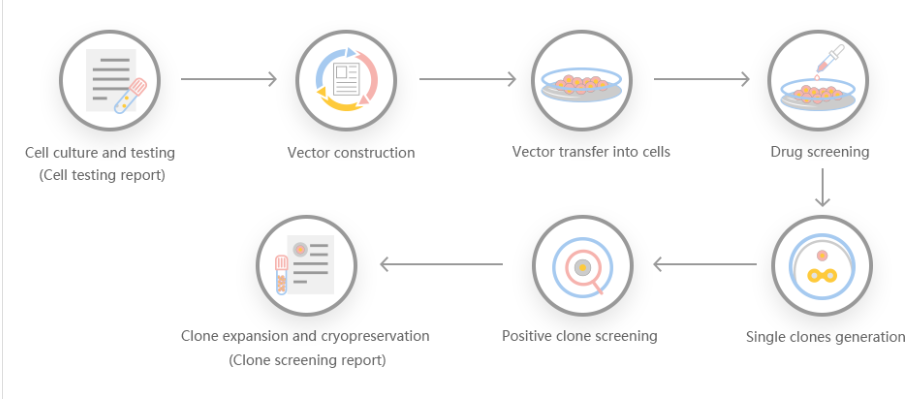
Genome editing and the creation of cellular models can advance research programs in the area of functional genomics, signaling pathways, metabolism, cell death, drug discovery, drug response, and cancer research, etc. The CRISPR system is a precise genome editing technique, which can create gene knockout or knock-in genome manipulations through the substitution of a target genetic sequence with a desired donor sequence. H1299  is a model human lung adenocarcinoma cell line, used for lung cancer study mainly drug discovery, disease mechanisms, initiation, progression, and therapeutics. CRISPR-system mediated edited H1299 cell lines allow investigators to study cancer hallmarks, disclosure of drug resistance mechanism, cancer therapeutics, cell death research, functional genomics, signaling pathways, drug discovery, drug response, and cell therapy. CRISPR/Cas9 technology positively fuel the advancement of in vivo and in vitro gene editing in lung cancer and have an immense impact on molecular medicine. Besides, knockout of the RSF1 gene in combination with paclitaxel resulted in the cell-cycle arrest in G1, increased apoptosis, and reduced cell migration and proliferation. Also, Knocking out NESTIN in H1299 cells can promote apoptosis, inhibit proliferation and colony formation, and suppress cell invasion by inhibiting epithelial-to-mesenchymal transition (EMT). Ubigene developed CRISPR-U™ for gene manipulation of the H1299 cell line. Thereby, possible to achieve genome-edited cells with the utilization of the CRISPR/Cas9 system. Ubigene can customize the gene-editing in eukaryotic cells as well as can generate various genes modification in animal models.

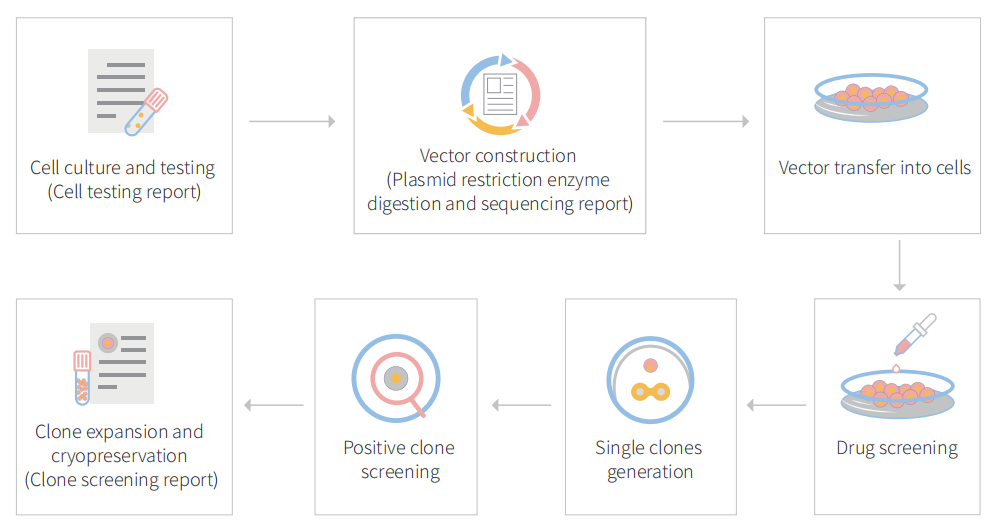
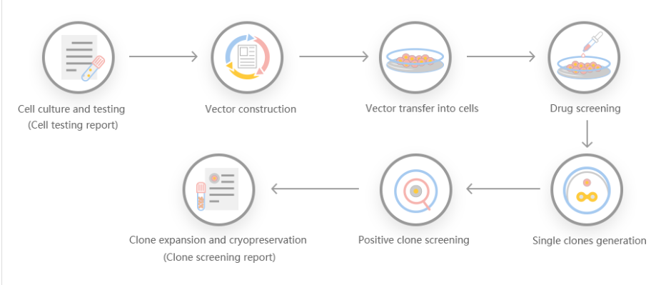
Figure: CRISPR-U™ customized workflow for engineered H1299 model cells

 Case study :

BCAR1 knockout cell line reveals that BCAR1 promotes proliferation and cell growth in lung adenocarcinoma via upregulation of POLR2A

Breast cancer antiestrogen resistance protein 1 (BCAR1; Crk‐associated substrate, CAS; p130cas) directly interact with the multiple protein motifs of various phosphatases and kinases, and to mediate Src through FAK bridge indirect association. BCAR1 has been reported to enhance tumor proliferation, invasion, and metastasis in several cancers (prostate cancer, endometrial adenocarcinoma, oral squamous cell carcinoma, breast cancer, and lung cancer). it also predicts poorer prognosis in lung adenocarcinoma cases. 


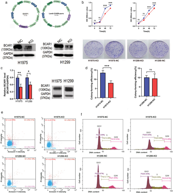
In this study, the researcher explores the role of BCAR1 in proliferation and cell growth in lung adenocarcinoma and the networks of proteins that interact with BCAR1 to trigger the proliferation of lung cancer cells. They used to lung adenocarcinoma cell line (NCl-H1975 and NCl-H1299) and established BCAR1 knockout cell line by CRISPR/Cas9 system as well as performed KO cell line for cell proliferation, colony formation, apoptosis, and cell cycle assay (Fig.1) They found that BCAR1 expression in the KO group was significantly lower than that of control as well as proliferation was significantly inhibited (Fig.1 b,c). Colony‐formation efficiency of H1975 cells was significantly decreased but no significant difference between BCAR‐KO and NC in H1299 cells (Fig.1d). Apoptosis or cell cycle progression analysis revealed that no difference in apoptosis and cell cycle of H1975 and H1299 cells following BCAR‐KO (Fig.1 e,f). 

Figure 1: Cell proliferation, colony formation, apoptosis, and cell cycles of H1975 and H1299 cell after BCAR1 knockout


They overexpressed BCAR1 in 293T cell lines and performed immune precipitation-mass spectrometric (IP-MS) as well as different downstream analysis. The IP-MS and bioinformatic analysis predict potential interactions with BCAR1 (Fig. 2). Bioinformatic analysis revealed potential BCAR1 interact proteins and interaction partners catalytic activity and transferase activity (Fig.2a,b). The Cancer Genome Atlas (TCGA) database verification confirms BCAR1 overexpression correlates with cancer (Fig.2c). They analyze cancer-related interacting partner PPI analysis and found that high expression of POLR2A, MAPK3, MOV10, and XAB2 predicted poor prognosis in lung adenocarcinoma (Fig.2d). They consider POLR2A for further verification due to the link with more genes and the possibility of involvement in the signaling cascade.


Figure-2: Bioinformatic analysis of BCAR1 overexpressed in 293cell line


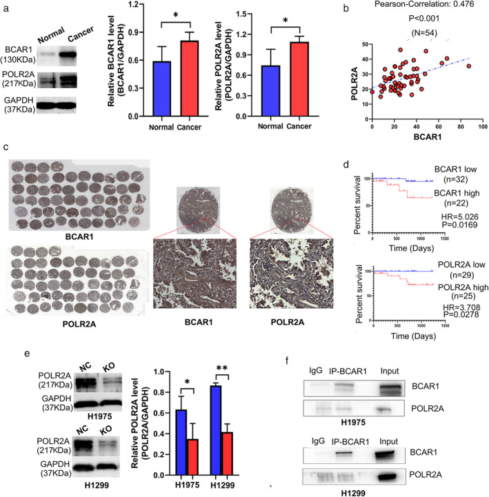
POLR2A and BCAR1 were significantly increased in lung adenocarcinoma tissues compared to adjacent normal tissues (Fig.3a). IHC‐stained TMA is shown in Fig 3b, demonstrate that BCAR1 was expressed in the nucleus, in the cytoplasm, or both locations (Fig.3c). However, POLR2A was highly expressed in the nucleus. POLR2A expression was significantly positively correlated to BCAR1 expression (R = 0.476, P < 0.001). Neither BCAR1 nor POLR2A expression was correlated with tumor size. High expression of either BCAR1 or POLR2A predicted poor prognosis in 54 lung cancer cases in the early-stage (Fig. 3d). They further verified POLR2A in H1975, and H1299 cell KO clone and western blot analysis demonstrated that POLR2A was significantly decreased in BCAR1‐KO compared to NC cells (Fig.3e). BCAR1 regulates POLR2A in H1975 and H1299 cells, despite the negative results of the CO‐IP (Fig.3f).


Figure-3: The relationship between BCAR1 and POLR2A as well as their prognostic significance in lung adenocarcinoma.


In summary, the researcher did not find a direct interaction between BCAR1 and POLR2A in H1975 and H1299 cells. The underlying mechanism of how BCAR1 and POLR2A are connected remains unclear. Moreover, synergism analysis is needed to demonstrate the role of interaction between BCAR1 and its partners concerning proliferation and cell growth in lung cancer. Future robust studies are required to resolve the interesting abovementioned points. 


CRISPR Gene-Editing Breast Cancer Treatment might Change the Face of Modern Medicine|Ubigene


The murine mammary carcinoma cell line 4T1 is widely used as a syngeneic tumor model for human breast cancer, a tumor entity with the world-wide highest incidence. 4T1 cells can easily be transplanted into the mammary gland and they are poorly immunogenic, highly tumorigenic, invasive, spontaneously metastasizing to distant organs. Thus, the location of the primary tumor and its metastatic spreading closely resemble the clinical course in patients. Moreover, 4T1 cells are used to investigate triple-negative breast cancer (TNBC) lacking protein expression of estrogen receptor (ER), progesterone receptor (PgR), and epidermal growth factor receptor 2 (ErbB2). This triple-negative phenotype is estimated for more than 17% of breast cancers that are annually diagnosed. Earlier studies reported that mice vaccinated with an EMT6 vaccine exhibited significantly greater protective immunity than mice vaccinated with a 4T1 vaccine. Hybrid vaccination studies revealed that the 4T1 vaccination-induced both local and systemic immune impairments. Thus, 4T1 is among the most popular syngeneic tumor models used for assessing novel therapeutic approaches.


Breast cancer is the second most common cause of death in women in the United States. Most breast cancer deaths are due to metastasis to distal sites of recurrence at the primary site. Breast cancer typically metastasizes to the lung, liver, brain, lymph nodes, and bone. Since one model will not fit all studies, depending on the nature of the study, many murine models are used to mimic the natural process of breast cancer. These models include the tissue or cell inoculation, via ectopic, xenograft, subcutaneous, orthotopic, intra-cardiac.


Researcher choice 4T1 cell line has several characteristics that make it suitable for experiments in the cellular model as well as in animal models for human cancer. It has several advantages over other cancer cell lines because it closely resembles tumor growth and metastasis as seen in human breast cancers. To date, most research on genome editing is designed to better understand diseases by using cultured cells and animal models, while scientists work to gauge the safety and effectiveness of these approaches for human use. Genome editing, and biomarker testing, open the door to patient-tailored therapies that will change outcomes and save lives. This is the future of breast cancer care. CRISPR/Cas9 genome-editing technology has brought invaluable opportunities in the field of breast cancer gene/ cell therapy and could have a crucial role in breast cancer treatment.

 Application:

1. Multi-omics studies: genomics, transcriptomics, proteomics, and immunome.

2. Cancer recurrence: Identify the cancer recurrence-associated genes, interaction network, downstream studies, and preventive measures of tumor recurrence.

3. Gene discovery: Identify new cancer driver genes and uncover cancer-specific vulnerabilities.

4. Cancer vaccine: Autologous tumor cell vaccines (ATCVs), Tumor-derived cytokine, ATCV immunogenicity, immunosuppressive cytokines, cancer cell-based vaccines, etc. Study on genetically modified tumor cells for therapeutic cancer vaccine or preventive cancer vaccine.

5. Drug discovery: Novel therapeutic vulnerabilities and the development of more effective treatments

6. Disease modeling: Allograft and xenograft disease modeling, disease progression, and mechanism studies.

  CRISPR-U™ gene editing in 4T1 cells

Genome editing is the process of precisely modifying the nucleotide sequence within a gene for wiping out or initiating explicit and preferred characters in the genome. CRISPR/Cas9 can correct errors in the genome and switch on or off certain genes in cells and organisms fast, cheaply, and relatively easily. 4T1 cell is a model murine carcinoma cell line and is broadly used as syngeneic tumor models for human breast cancer study (drug-resistant gene, chemotherapy-resistant gene, drug discovery, disease mechanisms, initiation, progression, and therapeutics). Gene editing within this cell line possible to generate single or multiple gene knockouts, mutation corrections, or insert reporter transgenes. These genome-edited 4T1 cell lines will allow investigators to study breast cancer hallmarks, disclosure of drug resistance mechanism, cancer therapeutics, cell death research, functional genomics, signaling pathways, drug discovery, drug response, and cell therapy. CRISPR/Cas9 technology positively fuel the advancement of in vivo and in vitro gene editing in breast cancer to tackle the complexity of breast cancer metastasis, drug resistance, chemotherapy resistance and have an immense impact on precision medicine. Ubigene developed CRISPR-U™ for gene manipulation of the 4T1 cell line. Thereby, possible to achieve genome-edited cells with the utilization of the CRISPR/Cas9 system. Ubigene can customize the gene-editing in eukaryotic cells as well as can generate various genes modification in animal models.


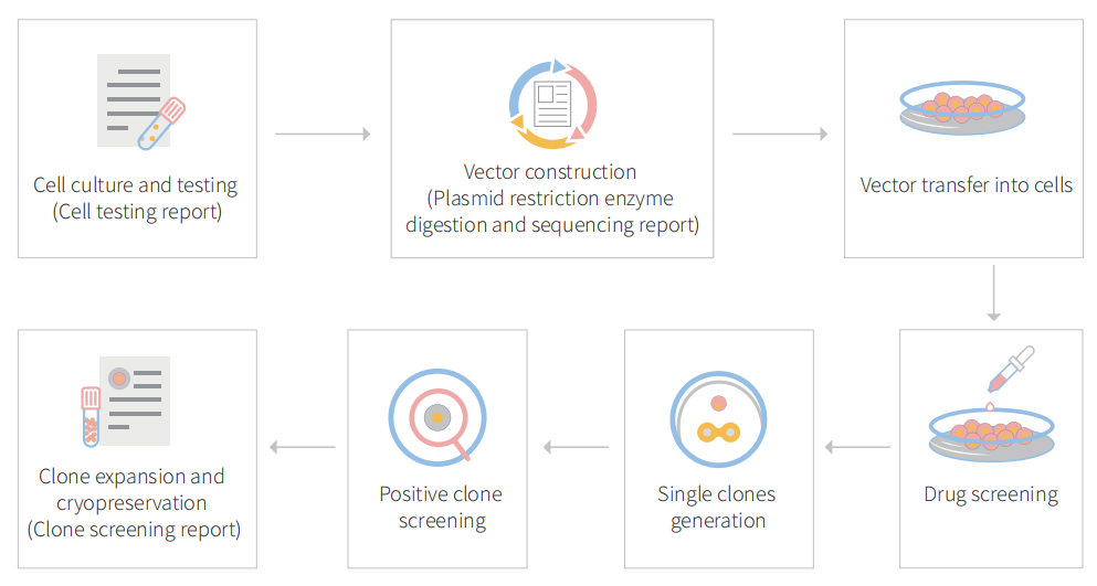
CRISPR-U™ customized workflow for engineered 4T1 model cells

Figure: CRISPR-U™ customized workflow for engineered CT26.WT  model cells

 Case study :

CRISPR/Cas9 mediated breast cancer research shows endogenous PAD4 promotes lung metastasis by extracellular chromatin formation

Breast cancer is the most common cancer and the second leading cause of cancer deaths in women in the United States. Along with the aggressive growth of primary tumor mass, the development of distant metastases is a major cause of death from breast cancer. Due to the heterogeneity and lack of well-defined molecular targets, patients with metastatic breast cancer have a poor 5-year survival rate, and finding a method to limit metastasis remains a great challenge. Pathology studies detected the significant expression of PAD4 in many tumor tissues derived from the colon, duodenum, esophagus, fallopian tube, gall bladder, lung, ovary, parotid, pancreas, prostate, rectum, small intestine, stomach, thyroid, and uterus. Peptidyl Arginine Deiminase 4 (PAD4/PADI4) is a posttranslational modification enzyme that converts protein arginine or mono-methylarginine to citrulline.


Overview of PAD4 mediated Lung metastasis

Overview of PAD4 mediated Lung metastasis

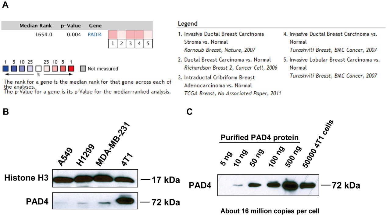
To determine whether PAD4 expression was associated with breast cancer in humans, the researcher interrogated the Tissue Cancer Genome Atlas and Oncomine database. After a meta-analysis of gene expression profiling, they found that PAD4 elevated expression was significantly associated with breast carcinoma compared with normal (Fig. 1A). To investigate the functional role of PAD4 in breast cancer, they found highly metastatic mammary carcinoma 4T1 cells express a much higher level of PAD4 than other cancer cell lines (Fig. 1B). They quantify the amount of PADI4 in 4T1 cells and found that PADI4 protein from 5 × 10 4 4T1 cells is roughly comparable to 100 ng purified PAD4 protein, which corresponds to ~16 million molecules of PAD4 protein per cell (Fig. 1C).


 PAD4 expression in human and mouse mammary tumors.

Fig 1: PAD4 expression in human and mouse mammary tumors.

PAD4 is a calcium-dependent enzyme. Therefore, they performed Western blot experiments after calcium ionophore treatment and found that histone H3 citrullination detected with the H3Cit antibody (made against a histone H3 N-terminal peptide containing Cit2, Cit8, and Cit17) increased significantly after calcium ionophore treatment (Fig. 2A). To test how histone citrullination activity affects chromatin structure they analyzed cell morphology with immunostaining, H3Cit staining was greatly increased at areas of highly decondensed chromatin (Fig. 2B). They tested the citrullination level on two positions with α-H3Cit26 (Fig. 2C and 2D) and α-H4Cit3 antibodies (Fig. 2E and 2F) respectively. To further confirm the presence of extracellular chromatin structure, intact 4T1 cells with or without calcium ionophore treatment were subjected to digestion with MNase, a nuclease that cuts between nucleosomes in chromatin (Fig. 2G). Accordingly, histone H3 protein can only be detected from the supernatant of 4T1 cells after calcium ionophore stimulation (Fig. 2H). Together, they found a modest increase in extracellular DNA and H3Cit staining after exposure of 4T1 cells to HMGB1, indicating the ability of HMGB1 to induce CECN in 4T1 cells (Fig. 2I and 2J).


Activation of PADI4 leads to hyper-citrullination and CECN formation in 4T1 cells in-vitro.

Fig 2: Activation of PADI4 leads to hyper-citrullination and CECN formation in 4T1 cells in-vitro.

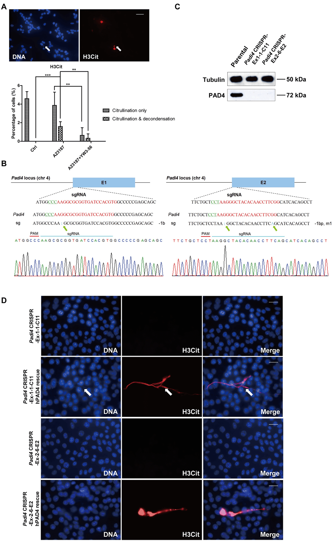
They found that inhibition of PAD decreased the incidence of both hyper-citrullination and release of CECN (Fig. 3A). They knocked out Padi4 in tumor cells using the CRISPR/Cas9 method and selected two single-cell colonies named Padi4 CRISPR-Ex1–1-C11 and Padi4 CRISPR-Ex2–6-E2 with Padi4 deletion in exon 1 and 2, respectively. The deletion of Padi4 was confirmed by Sanger Sequencing, and Western blot (Fig. 3B, C). Citrullination and the formation of CECN were not detected after calcium ionophore stimulation (Fig. 3D). Thus, PAD4 activity is required for histone hyper-citrullination and CECN formation.


Formation of CECN is dependent on PAD4.

Fig 3: Formation of CECN is dependent on PAD4.

To investigate whether 4T1 cells can release CECN in vivo, they compared citrullination levels from orthotopically transplanted and size-matched 4T1 wild type and Padi4 knockout tumors. PAD4 was only detected in the parental cell line and parental tumors (Fig. 4A). To test whether hyper-citrullination leads to decondensed chromatin are shown in Fig. 4B. Immunostained were performed to detect potential CECN structure (Fig. 4C). H&E staining and immunostaining experiments with tumors allografted on BALB/c Padi4 knockout mice are shown in Fig. 4D to 4F. The H3Cit staining in the primary tumor is area-based and has different staining patterns, with some areas positive while others negative of H3Cit staining (Fig. 4G). This is likely due to the heterogeneity of tumor microenvironment with different levels of ROS or numbers of dying cells.


CECN is present in allograft tumors.

Fig 4: CECN is present in allograft tumors.

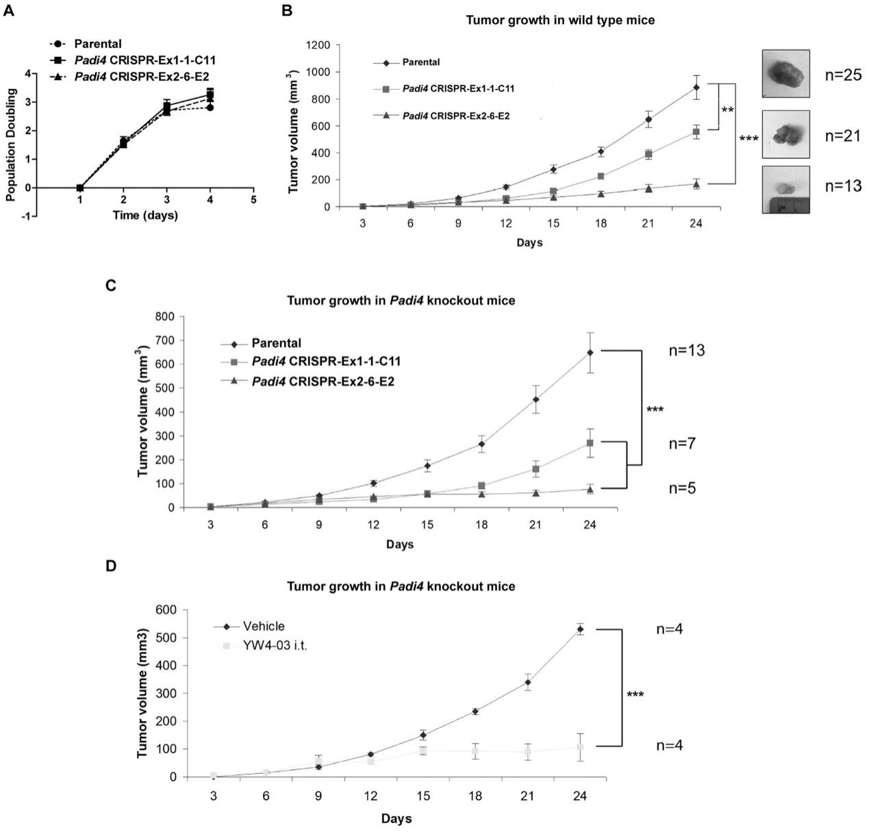
To assess the cell-autonomous role of PAD4 on tumor growth and metastasis, wild type female BALB/c mice were inoculated in the mammary gland with 4T1 wild type and Padi4 knockout cells. Although all 4T1 cell types showed similar degrees of in vitro growth rates (Fig. 5A), the allograft Padi4 knockout tumors grew significantly slower compared with wild type 4T1 tumors in wild type BALB/c mice (Fig. 5B). The reduced growth rate of Padi4 knockout tumors was also observed in Padi4 knockout BALB/c mice (Fig. 5C). Similarly, intratumoral PAD4 inhibitor treatment also reduced the primary tumor growth rate (Fig. 5D).


Growth rates of primary mammary tumors formed by 4T1 cells with different Padi4 genotypes or combined with YW4–03 treatment.

Fig 5: Growth rates of primary mammary tumors formed by 4T1 cells with different Padi4 genotypes or combined with YW4–03 treatment.

4T1 cells are highly metastatic compared with a sibling cell line 67NR which was derived from the same mouse tumor but cannot metastasize. As shown in Fig. 6A, the PAD4 level is significantly higher in 4T1 cells compared with 67NR cells. After allograft, mice bearing wild type tumors formed a significantly higher number of metastatic foci on the lung surface than the two Padi4 knockout cell lines (Fig. 6B and 6C). Lung metastasis was still significantly reduced in mice bearing tumors derived from Padi4 CRISPR cancer cells even after prolonged growth (Fig. 6D–6F). To test the effects of PADI4 inhibition on a population level, lung metastasis was also compared in the YW4–03 intratumoral treatment model using Padi4 knockout mice (Fig. 6G and 6H).


 Lack of PAD4 in tumors leads to reduced lung metastasis.

Fig 6: Lack of PAD4 in tumors leads to reduced lung metastasis.

To further test the idea that CECNs are involved in metastasis, they used DNase I to disrupt extracellular DNA including CECN structures in the 4T1 wild-type tumors growing in the Padi4 knockout mice, lung metastasis was significantly decreased (Fig. 7A and 7B). Also, tested whether PAD4 expressed in cancer cells influences intravasation by analyzing the Circulating Tumor Cells (CTCs) in blood with colony formation assays. Neither loss of Padi4 nor DNase I treatment influenced the CTC numbers from size-matched tumor-bearing mice, suggesting cancer cell endogenous PAD4 does not promote metastasis through the intravasation process (Fig. 7C–7F). To further test whether PADI4 is functioning at steps after intravasation, they performed experimental metastasis assays by intravenously injecting the same number of tumor cells into mice. The loss of Padi4 in 4T1 cells didn’t significantly reduce the number of cancer cells accumulated compared with the Padi4 wild type cells (Fig. 7G and 7H).


Cancer cell endogenous PAD4 promotes metastasis through steps after intravasation.

Fig 7: Cancer cell endogenous PAD4 promotes metastasis through steps after intravasation.

In summary, the researcher establishes the occurrence of PADI4-mediated CECN in 4T1 cells in vitro and in vivo. The tumor microenvironment harbors the signal to activate PADI4 and deletion of Padi4 or removal of extracellular DNA including CECN with DNase I in 4T1 cells inhibits metastatic lung colonization. PAD4’s vital role in promoting tumor growth and metastasis renders itself a tangible therapeutic.

Ubigene developed CRISPR-U™ which optimizes eukaryotic cells and animal gene-


editing vectors and processes. The efficiency and accuracy are 10x higher than traditional methods. Contact us immediately to know about your research related services!


Monday, November 30, 2020


Saccharomyces Cerevisiae Knockout Cell line | CRISPR


Saccharomyces cerevisiae is a species of yeast. It has been instrumental to winemaking, baking, and brewing since ancient times. It is believed to have been originally isolated from the skin of grapes (one can see the yeast as a component of the thin white film on the skins of some dark-colored fruits such as plums; it exists among the waxes of the cuticle). It is one of the most intensively studied eukaryotic model organisms in molecular and cell biology, much like Escherichia coli as the model bacterium. It is the microorganism behind the most common type of fermentation.


Phosphoglycerate mutase knock-out mutant Saccharomyces cerevisiae: physiological investigation and transcriptome analysis


The yeast Saccharomyces cerevisiae is able to adapt its metabolism to grow on different carbon sources and to shift to non-fermentative growth on C2 or C3 carbon sources (ethanol, acetate, or glycerol) through the activation of gluconeogenesis. Researcher studied the response to the deletion of the glycolytic and gluconeogenic gene GPM1, encoding for phosphoglycerate mutase. It was previously shown that a S. cerevisiae strain with non-functional copies of GPM1 can only grow when glycerol and ethanol are both present as carbon sources, whilst addition of glucose was shown to strongly inhibit growth. It was suggested that glycerol is needed to feed gluconeogenesis whilst ethanol is required for respiration. Researcher studied the physiological response of the GPM1 knock-out mutant through fermentation and transcriptome analysis. Furthermore, researcher compared the physiological results with those obtained through simulations using a genome-scale metabolic model, showing that glycerol is only needed in small amounts for growth. Our findings strongly suggest a severely impaired growth ability of the knock-out mutant, which presents increased transcript levels of genes involved in the pentose phosphate pathway and in the glyoxylate shunt. These results indicate an attempt to compensate for the energy imbalance caused by the deletion of the glycolytic/gluconeogenic gene within the mutant.


Reducing phenolic off-flavors through CRISPR-based gene editing of the FDC1 gene in Saccharomyces cerevisiae x Saccharomyces eubayanus hybrid lager beer yeasts


Today’s beer market is challenged by a decreasing consumption of traditional beer styles and an increasing consumption of specialty beers. In particular, lager-type beers (pilsner), characterized by their refreshing and unique aroma and taste, yet very uniform, struggle with their sales. The development of novel variants of the common lager yeast, the inter-specific hybrid Saccharomyces pastorianus, has been proposed as a possible solution to address the need of product diversification in lager beers. Previous efforts to generate new lager yeasts through hybridization of the ancestral parental species (S. cerevisiae and S. eubayanus) yielded strains with an aromatic profile distinct from the natural biodi-versity. Unfortunately, next to the desired properties, these novel yeasts also inherited unwanted characteristics. Most notably is their phenolic off-flavor (POF) production, which hampers their direct application in the industrial production processes. Researchers describe a CRISPR-based gene editing strategy that allows the systematic and meticulous intro-duction of a natural occurring mutation in the FDC1 gene of genetically complex industrial S. cerevisiae strains, S. eubayanus yeasts and interspecific hybrids. The resulting cis-genic POF- variants show great potential for industrial application and diversifying the cur-rent lager beer portfolio.


A CRISPR/Cas9-based exploration into the elusive mechanism for lactate export in Saccharomyces cerevisiae.


CRISPR/Cas9-based genome editing allows rapid, simultaneous modification of multiple genetic loci in Saccharomyces cerevisiae. This technique was used in a functional analysis study aimed at identifying the hitherto unknown mechanism of lactate export in this yeast. First, an S. cerevisiae strain was constructed with deletions in 25 genes encoding transport proteins, including the complete aqua(glycero)porin family and all known carboxylic acid transporters. The 25-deletion strain was then transformed with an expression cassette for Lactobacillus casei lactate dehydrogenase (LcLDH). In anaerobic, glucose-grown batch cultures this strain exhibited a lower specific growth rate (0.15 vs. 0.25 h−1) and biomass-specific lactate production rate (0.7 vs. 2.4 mmol g biomass−1 h−1) than an LcLDH-expressing reference strain. However, a comparison of the two strains in anaerobic glucose-limited chemostat cultures (dilution rate 0.10 h−1) showed identical lactate production rates. These results indicate that, although deletion of the 25 transporter genes affected the maximum specific growth rate, it did not impact lactate export rates when analysed at a fixed specific growth rate. The 25-deletion strain provides a first step towards a ‘minimal transportome’ yeast platform, which can be applied for functional analysis of specific (heterologous) transport proteins as well as for evaluation of metabolic engineering strategies.

The efficiency of gene knock-out and cleavage can not only give people the ability to generate protein radical profiles and establish regulatory records, but also has many advantages, making it a particularly attractive recombinant protein expression system. First, it is carboxylated on glutamic acid and sulfated on tyrosine. Second, the operation is simple, and the recombinant protein can be quickly produced through transient gene expression. Third, it can be used for stable recombinant protein production. Some researchers used gene cell knockout and cutting efficiency systems to generate gene-edited cell lines, targeted sequencing of GLUL genomic loci, produced stable cell lines, and discovered the mechanism of stable expression of recombinant erythropoietin in humans .


According to customer needs, Yuanjing Biotechnology designs a stable gene transfer knockout program based on the target gene.


Scheme 1: Small-segment gene knockout program, gRNA is set in the introns at both ends of exon 2, and the number of bases encoded by the knockout exon is not 3 times, and the knockout can cause frameshift.
Scheme 2: Frameshift gene knockout scheme, gRNA is set on the exon, the number of missing bases is not 3 times, and frameshift mutation can occur after knockout.
Scheme 3: Large-segment gene knockout scheme, knock out the coding sequence of the entire gene to achieve the effect of large-segment knockout.


Reference

Papini M , Nookaew I , Scalcinati G , et al. Phosphoglycerate mutase knock-out mutant Saccharomyces cerevisiae: Physiological investigation and transcriptome analysis[J]. Biotechnology Journal, 2010, 5(10):1016-1027.

Mertens S , Gallone B , Steensels J , et al. Reducing phenolic off-flavors through CRISPR-based gene editing of the FDC1 gene in Saccharomyces cerevisiae x Saccharomyces eubayanus hybrid lager beer yeasts[J]. Plos One, 2019, 14(1).

Robert, Mans, Else-Jasmijn, et al. A CRISPR/Cas9-based exploration into the elusive mechanism for lactate export in Saccharomyces cerevisiae.[J]. FEMS yeast research, 2017.


Ubigene Biosciences is co-founded by biological academics and elites from China, the United States, and France. We are located in Guangzhou Science City, which serves as a global center for high technology and innovation. Ubigene Biosciences has 1000㎡ office areas and laboratories, involving genome editing, cell biology technology, and zebrafish research. We provide products and services for plasmids, viruses, cells, and zebrafish. We aim to provide customers with better gene-editing tools for cell or animal research.

We developed CRISPR-U™ and CRISPR-B™(based on CRISPR/Cas9 technology) which is more efficient than general CRISPR/Cas9 in double-strand breaking, CRISPR-U™ and CRISPR-B™ can greatly improve the efficiency of homologous recombination, easily achieve knockout (KO), point mutation (PM) and knockin (KI) in vitro and in vivo. 

Genome Editing Platform
——Focusing on the Application of CRISPR-U™ and CRISPR-B™ Gene Editing Technology
1. Provides various types of gene-editing vectors for different species.
2. Provides different virus packaging services, including lentiviruses, adenoviruses and adeno-associated viruses.3. Provides high-quality services for gene knockout, point mutation and knockin cell lines. 

Cell Biology Platform
——Focusing on primary cell
1. Provides over 400 types of primary cells.
2. Provides culture strategies and related products for different cell types.3. Provides cell biology-related services such as cell isolation, extraction and validation.

    Friday, August 7, 2020

    Multi Crispr Knockout Bacteria

    Scientists have developed a continuous knockout cell line technology based on the CRISPR-Cas9 system, including single and multiple (up to three) target gene insertions and knockouts for the purpose of bacterial modification. The CRISPR/Cas system is a kind of prokaryotic immunity. The resistance of the system to foreign genetic elements (such as those found in plasmids and phages) can provide some form of acquired immunity. RNA with spacer sequence can help Cas protein recognize and cleave foreign DNA. Other Cas proteins guided by RNA can cleave foreign RNA. CRISPR is found in approximately 40% of bacterial genome sequences and 90% of archaeal sequences.

    Multi-gene editing in the E. coli genome through the CRISPR-Cas9 system

    The construction of industrially useful microorganisms requires effective genome-scale editing tools. The researchers describe a targeted, continuous multi-gene editing strategy that uses the Streptococcus pyogenes type II CRISPR-Cas9 system to be applied to the E. coli genome to achieve a variety of precise genome modifications, including gene deletion and insertion, with efficiency The maximum is 100%, and it can perform multi-gene editing on three targets at the same time. The system also proved that it successfully achieved targeted chromosome deletion in another Enterobacteriaceae-Tatumella citrea, with an efficiency of up to 100%.

    Multi-stage knockout of Escherichia coli using CRISPR/Cas9

    With the recent use of CRISPR/Cas9 technology as a standard tool for genome editing, laboratories around the world have experienced one of the largest molecular biology advances since PCR. The main advantage of this method is its simplicity and versatility for any category. Of particular interest is the widely studied Gram-negative bacterium Escherichia coli, because it is considered a major force for research and industrial applications. The researchers proposed a simple, reliable and effective scheme using the CRISPR/Cas9 system combined with the lambda Red machine for gene knockout in E. coli. In our procedure, it is crucial to use double-stranded donor DNA and a solidification strategy to remove the RNA-encoding guide plasmid, which allows the initiation of new mutations in only two working days. Our protocol allows multiple stepwise knockout strains with high mutagenesis efficiency to be suitable for high-throughput methods.

    Multi-functional genome-wide CRISPR system for high throughput genotype–phenotype mapping

    Genome-scale engineering is an indispensable tool to understand genome functions due to our limited knowledge of cellular networks. Unfortunately, most existing methods for genome-wide genotype–phenotype mapping are limited to a single mode of genomic alteration, i.e. overexpression, repression, or deletion. Researcher report a multi-functional genome-wide CRISPR (MAGIC) system to precisely control the expression level of defined genes to desired levels throughout the whole genome. By combining the tri-functional CRISPR system and array-synthesized oligo pools, MAGIC is used to create, to the best of our knowledge, one of the most comprehensive and diversified genomic libraries in yeast ever reported. The power of MAGIC is demonstrated by the identification of previously uncharacterized genetic determinants of complex phenotypes, particularly those having synergistic interactions when perturbed to different expression levels. MAGIC represents a powerful synthetic biology tool to investigate fundamental biological questions as well as engineer complex phenotypes for biotechnological applications.

    Ubigene developed CRISPR-B™ which optimizes the microbial gene-editing vectors and process. The efficiency and accuracy are much higher than traditional methods. CRISPR-B™ can be used in gene editing of bacteria and fungi

    Reference

    Endo M, Mikami M, Toki S. Multigene knockout utilizing off-target mutations of the CRISPR/Cas9 system in rice. Plant Cell Physiol. 2015;56(1):41-47. doi:10.1093/pcp/pcu154

    König, Enrico & Zerbini, Francesca & Zanella, Ilaria & Fraccascia, Davide & Grandi, Guido. (2018). Multiple Stepwise Gene Knockout Using CRISPR/Cas9 in Escherichia coli. BIO-PROTOCOL. 7. 10.21769/BioProtoc.2688.

    Jiazhang Lian, Carl Schultz, Mingfeng Cao, Mohammad HamediRad,Huimin Zhao.Multi-functional genome-wide CRISPR system for high throughput genotype–phenotype mapping.Nature Communications .2019.

    The efficiency of gene knock-out and cleavage can not only give people the ability to generate protein radical profiles and establish regulatory records, but also has many advantages, making it a particularly attractive recombinant protein expression system. First, it is carboxylated on glutamic acid and sulfated on tyrosine. Second, the operation is simple, and the recombinant protein can be quickly produced through transient gene expression. Third, it can be used for stable recombinant protein production. Some researchers used gene cell knockout and cutting efficiency systems to generate gene-edited cell lines, targeted sequencing of GLUL genomic loci, produced stable EPO cell lines, and discovered the mechanism of stable expression of recombinant erythropoietin in humans .

    According to customer needs, Yuanjing Biotechnology designs a stable gene transfer knockout program based on the target gene.
    Scheme 1: Small-segment gene knockout program, gRNA is set in the introns at both ends of exon 2, and the number of bases encoded by the knockout exon is not 3 times, and the knockout can cause frameshift.
    Scheme 2: Frameshift gene knockout scheme, gRNA is set on the exon, the number of missing bases is not 3 times, and frameshift mutation can occur after knockout.
    Scheme 3: Large-segment gene knockout scheme, knock out the coding sequence of the entire gene to achieve the effect of large-segment knockout.

    Ubigene Biosciences is co-founded by biological academics and elites from China, the United States, and France. We are located in Guangzhou Science City, which serves as a global center for high technology and innovation. Ubigene Biosciences has 1000㎡ office areas and laboratories, involving genome editing, cell biology technology, and zebrafish research. We provide products and services for plasmids, viruses, cells, and zebrafish. We aim to provide customers with better gene-editing tools for cell or animal research.

    We developed CRISPR-U™ and CRISPR-B™(based on CRISPR/Cas9 technology) which is more efficient than general CRISPR/Cas9 in double-strand breaking, CRISPR-U™ and CRISPR-B™ can greatly improve the efficiency of homologous recombination, easily achieve knockout (KO), point mutation (PM) and knockin (KI) in vitro and in vivo. 

    Genome Editing Platform
    ——Focusing on the Application of CRISPR-U™ and CRISPR-B™ Gene Editing Technology
    1. Provides various types of gene-editing vectors for different species.
    2. Provides different virus packaging services, including lentiviruses, adenoviruses and adeno-associated viruses.3. Provides high-quality services for gene knockout, point mutation and knockin cell lines. 

    Cell Biology Platform
    ——Focusing on primary cell
    1. Provides over 400 types of primary cells.
    2. Provides culture strategies and related products for different cell types.3. Provides cell biology-related services such as cell isolation, extraction and validation.

    [Research highlight] Enhancing p53 pathway can efficiently suppress colon cancer

      Colorectal cancer is the third most diagnosed cancer and leads to the second mortality among cancers worldwide. The first-line chemotherap...