Monday, February 7, 2022

[Research highlight] Enhancing p53 pathway can efficiently suppress colon cancer


Colorectal cancer is the third most diagnosed cancer and leads to the second mortality among cancers worldwide. The first-line chemotherapeutic drugs of colorectal cancer are mainly 5-fluorouracil(5-FU) based. However, these drugs exhibit compromised efficacy due to significant toxicity, drug resistance or patient inconvenienceNovel chemotherapeutic drugs for efficient treatment of colorectal cancer patients are urgently needed. Cabazitaxel (CBT), a microtuble inhibitor, can promotes polymerization of tubulin and stabilize microtubules. It shows antitumor activity in docetaxel-refractory metastatic prostate cancer and breast cancer.

However, it is not clear whether CBT is effective in inhibiting colorectal cancer and what the underlying mechanism is. In response to this question, The researchers from The Sixth Affiliated Hospital of Sun Yat-sen University, Wen Zhang et al. published an article named Cabazitaxel suppresses colorectal cancer cell growth via enhancing the p53 anti-tumor pathway” in FEBS Open Bio. In this study, the researchers screened 160 FDA approved drugs with HCT116 cells, the commonly used colorectal cancer cell lines, and found that Cabazitaxel, by enhancing the P53 antitumor pathway, can efficiently inhibit the proliferation and migration of colorectal cancer cells via a series of in vitro assays. 


Cabazitaxel inhibits colorectal cancer cell growth via activating p53 signaling pathway

KEGG pathway analysis revealed that CBT treatment induced genes were enriched in the well-known anti-tumor p53 signaling pathway (Fig. 1). Further gene set enrichment analysis (GSEA) revealed a positive correlation between p53 pathway genes with CBT upregulated genes in HCT116 cells, indicating that CBT indeed enhances the expression of p53 pathway genes (Fig. 2). To examine whether activation of p53 pathway plays a key role for CBT efficacy, they used CRISPR/Cas9 system to knock out TP53, p53 encoding gene, in HCT116 cells by two different gRNAs and generate TP53 KO1 cells and TP53 KO2 cells with two hTP53 gRNA KO plasmids (YKO-RP003-hTP53, YKO-RP003-hTP53) which were provided by Ubigene. MTT assay revealed that IC50 of CBT to TP53 KO1 cells and TP53 KO2 cells were about at least three fold higher than IC50 of CBT in HCT116 cells. The enhanced resistance to CBT of TP53 KO cells indicates that the inhibitory effect of CBT to HCT116 cells relies on the TP53 pathway (Fig. 3). All these results substantiate that CBT inhibits HCT116 cells mainly via activating p53 pathway. 

In this study, Ubigene provided hTP53 gRNA KO plasmids for the construction of TP53 knockout cell line. It was demonstrated that CBT inhibited HCT116 cells mainly by activating p53 pathway. Ubigene now provides TP53 knockout cell lines, which can be delivered within one week for only $1980. Click to learn more > >  

If gRNA plasmid is needed, welcome to visit our gRNA plasmid bank for retrieval. Click for more details > >  


Fig. 1
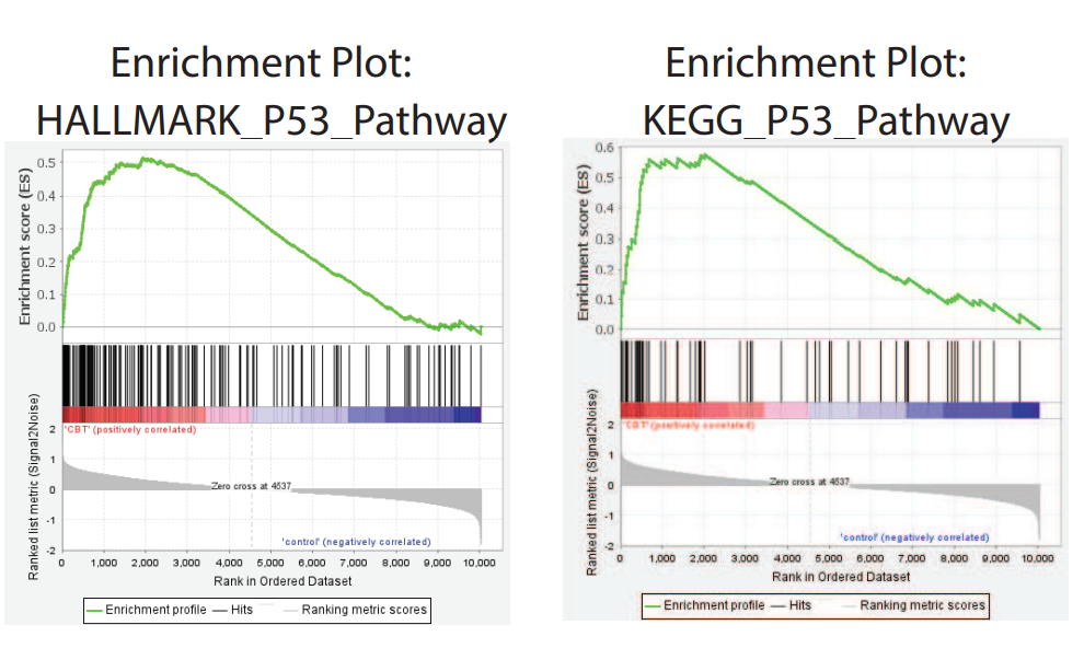
 

Fig. 2
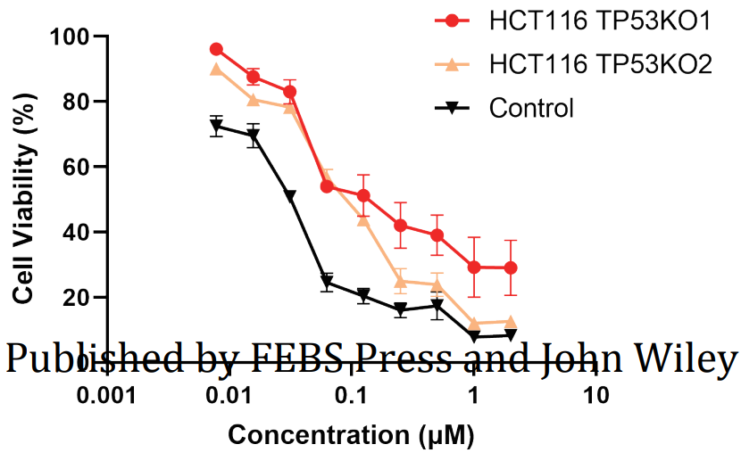
 

Fig. 3


In this study, they showed that CBT can efficiently inhibit colorectal cancer proliferation and migration.  It suppresses colorectal cancer via enhancing the expression of multiple p53 downstream effector genes and promoting cell cycle arrest, apoptosis and inhibition of angiogenesis. Hence, CBT may be served as an alternative option for colorectal cancer treatment in future. 


Ubigene is mainly focusing on cell engineering. We provide high-quality gene-editing cell lines, stable cell lines, virus packaging and other related services around the world, as well as nearly 2000 KO cell bank, gene knockout kits and other gene-editing related products.


More articles:

Well-known SW480 cell line is not “simple”

Gene editing in BEAS-2B cell line facilitates respiratory researches.

[Research Highlight] miR-302a is involved in vascular smooth muscle cell proliferation and restenosis


Vascular stenosis in different parts of body can lead to diseases of organs or tissues dominated by blood vessels in corresponding areas, such as Acute coronary syndrome(ACS) which is led by coronary artery stenosis and other cardiovascular diseases. ACS is a common serious cardiovascular disease, a serious type of coronary heart disease, percutaneous coronary interventions (e.g., balloon angioplasty or stenting) are considered the first-choice treatment for it. However, patients face an increased risk of restenosis after surgery. Excessive smooth muscle cell (SMC) proliferation, migration and invasion are considered important contributors to restenosis, which together lead to neointimal hyperplasia. Therefore, inhibition of SMC hyperplasia is an important therapeutic approach for restenosis.

MicroRNAs (miRNAs) are a family of endogenous noncoding small RNAs consisting of 1822 nucleotides that can regulate gene expression at the post-transcriptional level by pairing with imperfect complementary target sites in the 3UTR of their target mRNAs. Previous studies have suggested that miRNAs are involved in regulating SMC proliferation and migration. The miR-302a functions as a tumor suppressor by inhibiting Akt-dependent cell proliferation in different cancer cells, indicating that miR-302a may negatively regulate Akt activation. And the lack of Akt has been shown to inhibit VSMC proliferation and migration.

Given the importance of Akt in regulating cell proliferation, Ying-ying Liu et al from SUN YAT-SEN UNIVERSITY speculated that miR-302a is involved in Akt-mediated vascular SMC (VSMC) proliferation and restenosisThey published an article MicroRNA-302a promotes neointimal formation following carotid artery injury in mice by targeting PHLPP2 thus increasing Akt signaling” in Acta Pharmacologica Sinica. In the article, they used MiR-302 heterozygous mice and MiR-302 conditional KO mice building the carotid artery injury model, to investigated whether and how MicroRNAs regulated vascular SMC proliferation and vascular remodeling following carotid artery injury in mice. The study demonstrate that carotid artery injury-induced neointimal formation was remarkably ameliorated in miR-302 heterozygous mice and SMC-specific miR-302 knockout mice. In contrast, delivery of miR-302a adenovirus to the injured carotid artery enhanced neointimal formation. Upregulation of miR-302a enhanced the proliferation and migration of mouse aorta SMC (MASMC) in vitro by promoting cell cycle transition, whereas miR-302a inhibition caused the opposite results.


miR-302a potentiates MASMC proliferation by increasing Akt activity

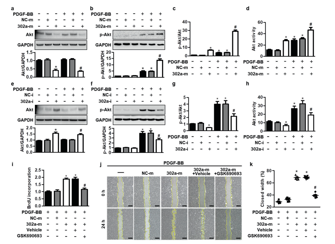
miR-302a promoted Akt activation by corporately decreasing Akt expression and increasing Akt phosphorylation in MASMCs. To further confirm whether Akt activity is required for the regulatory function of miR-302a in VSMC proliferation, cells were treated with miR-302a mimics and the Akt inhibitor GSK690693 in the presence of PDGF-BB. Compared with miR-302a mimics-treated MASMCs, treatment with GSK690693 inhibited cell proliferation and migration, which was potentiated by miR-302a upregulation. These results suggest that miR-302a regulates VSMC proliferation dependent on Akt signaling.

Figure 1


PHLPP2 is essential for the function of miR-302 in neointimal formation

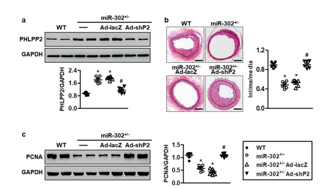
They further revealed that miR-302a directly targeted at the 3′ untranslated region of PH domain and leucine rich repeat protein phosphatase 2 (PHLPP2) and negatively regulated PHLPP2 expression. Restoration of PHLPP2 abrogated the effects of miR-302a on Akt activation and MASMC motility. To further evaluate the requirement of PHLPP2 during miR-302- regulated restenosis in vivo, recombinant adenovirus harboring leucine rich repeat protein phosphatase 2 shRNA (Ad-shPHLPP2) (provided by Ubigenewas administered to miR-302+/− mice. The result showed that the inhibitory effects of miR-302 knockdown on neointimal formation are largely dependent on the elevation of PHLPP2 expression(Fig.2).

The shRNA adenovirus provided by Ubigene in this study was used to further study the effect of PHLPP2 on the formation of mir-302 in neointima. The purity and titer of adenovirus determine its quality and way of use. The adenovirus particles provided by Ubigene are ultra-centrifuged and filtered, and the titer is determined by hexonimmunoassay to ensure the purity and accurate titer. At present, Ubigene provides adenovirus with titer of 10 ^ 10 ~ 10 ^ 12 PFU/ml. Click here to learn >>

Figure 2


In conclusion, the study demonstrated that miR-302a potentiates VSMC hyperplasia and subsequent neointimal formation by Akt activation via targeting its phosphatase PHLPP2, suggesting that inhibition of miR-302a may be a novel strategy for restenosis treatment, and this study demonstrated for the first time that PHLPP2 is a novel target of miR-302a. It provided a new idea for vascular restenosis treatment in future.


Ubigene is focusing on cell genome editing. We provide high-quality gene editing cell lines, stable cell lines, virus packaging and other related services. Our KO cell bank contains nearly 2000 KO cell lines, involving in 8 popular signal pathways, such as key genes of Akt signal pathway. 


More articles:

Versatile use of Luciferase cell line

Gold standard of biological production -- CHO cell line


[Research highlight] Enhancing p53 pathway can efficiently suppress colon cancer

  Colorectal cancer is the third most diagnosed cancer and leads to the second mortality among cancers worldwide. The first-line chemotherap...