Wednesday, March 31, 2021

Gene-Editing Salmonella: an ideal vector for tumor-targeting therapy | Ubigene

Salmonella is a facultative anaerobic gram-negative rod-shaped bacteria. Salmonella belongs to Enterobacteriaceae and is an important medical pathogen of humans and animals. Salmonella forms a complex bacterial community consisting of two species and six subspecies, including more than 2579 serotypes. At present, there are two species of Salmonella, S. entreica and S. bongori. The selective targeting of tumor tissue by Salmonella also makes it an ideal vector for tumorAs an intracellular parasite, Salmonella can effectively replicate and inhibit tumor growth in tumor tissue. After genetic engineering, it can be used as a carrier of tumor gene therapy in vitro and in vivo for liver cancer, gastric cancer and colorectal cancer.


 Salmonella and Tumor Therapy


1. Direct antitumor therapy with attenuated Salmonella


In view of the characteristics of Salmonella that can effectively inhibit tumor growth, scientists use a variety of genetic engineering techniques to modify the chromosome genome of Salmonella, which can reduce the virulence of Salmonella, so as to obtain the attenuated strain. While reducing the pathogenicity to the host, it still keeps high immunogenicity, thus ensuring the safety of clinical application.


2.Tumor targeting gene therapy


Attenuated Salmonella can carry exogenous genes, cytokines and exogenous effector proteins to treat tumors. Cytokines play an anti-tumor role by killing tumor cells directly. Exogenous effector proteins can be effectively transferred by attenuated Salmonella and expressed therapeutic proteins

 Knockout PhoP to construct attenuated Salmonella, improves the safety of   Salmonella in tumor treatment


The scientists used gene-splicing PCR combined with λ-Red system to delete PhoP from wild-type Salmonella typhimurium. phoP is a transcription regulator and a component of two regulatory systems that play key roles in adaptation to the homeostelium and in macrophage survival. It also controls the expression of more than 40 genes required by Salmonella typhimurium and its resistance to adverse environments in the host, such as low pH in the stomach, bile salt, hypoxia in the small intestine, and cationic antimicrobial peptides on epithelial cells. The disruption of phoP of Salmonella typhimurium leads to its inability to survive in phagocytes, and increases its sensitivity to stress, so as to achieve the function of reducing toxicity, and develop a safer bacterial therapy.


The phoP of wild type and standard strain of Salmonella typhimurium was disrupted by SOEing PCR. Three standard PCR and one fusion PCR were used to construct the linear DNA containing the upstream and downstream of phoP sequence and kanamycin cassette. pKD4 carrying kanamycin flanked by FRT (FLP recognition target) was used as a template plasmid. PCR confirmed that kanamycin had replaced phoP gene. Therefore, attenuated Salmonella was successfully constructed by removing phoP gene.


Gene-editing bacteria bring infinite possibilities to research, clinical medicine agriculture and other industrial productions!


Gene knockout has been used for many purposes, such as studying gene function, vaccine production and improving protein structure or expression. At present, the widely used traditional gene-editing methods include R6K suicide plasmid and λ-Red system.

Although the λ-Red system seems simple and has been successfully applied to E.coli and other gram-negative bacteria, the performance of this system in different bacteria is unstable due to the inherent differences of bacteria. In addition, suicide plasmid has some disadvantages such as host limitation and resistance residue. Other shortcomings of traditional KO include low recombination efficiency, heavy workload on validation, and residual loxP or FRT sites.


CRISPR/Cas9 technology is the most rapidly developed gene-editing technology in recent years. However, due to the lack of a repair system in bacteria, only a few microbial species were published to modify genes with this technology.Ubigene developed CRISPR-B™ technology, which is highly efficient in genome editing. CRISPR-B™ has features of easy-to-handle, accurate targeting, low off-target effect, scarless. It is efficient in various bacteria. The efficiency is more than 20x higher than that of traditional methods, easily achieve gene knockout, point mutation and knockin.

 A new treatment for tumor--CTNNB1-shRNA expression in Salmonella


Bacterial therapy has been used clinically for decades with a proven track record of safety in the treatment of gastroenteric diseases (for example, diarrhea, irritable bowel syndrome and inflammatory bowel disease). Moreover, recently there has been renewed interest in the clinical applications of live bacteria which is various non-pathogenic anaerobic bacteria are given intravenously and can infiltrate and replicate within solid tumors, particularly to treat human solid tumors. RNA interference (RNAi) has been established as an important research tool with great potential for gene therapy. Using a combination of bacterial therapy and RNAi therapy, scientists developed bacteria-mediated RNAi that delivers shRNA-expressing vectors to target cells to silence disease-causing genes.


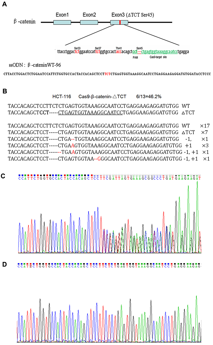
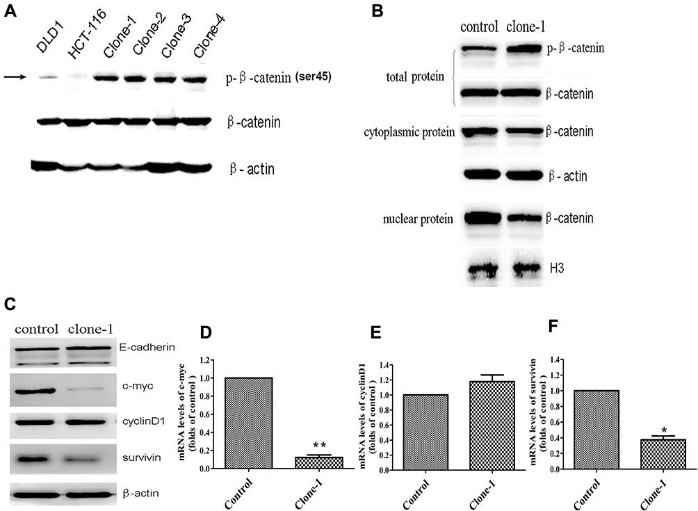
Scientists have established a pSLS plasmid system that can express shRNAs in attenuated Salmonella. shRNA against HIV tat gene, human and mouse CTNNB1 genes were inserted into the multiple cloning site of pSLS. Then these plasmids were transformed into attenuated Salmonella, resulting in corresponding SL-pSLS-TAT, SL-pSLS-huCAT and SL-pSLS-mCAT strains. To determine whether SL-PSLS-HucAT can knockdown CTNNB1 expression of SW480 and inhibit the growth of SW480, SW480 cells were treated at different MOI.


CTNNB1 gene silencing significantly reduced cell proliferation and death in SW480 cells compared with control cells.To determine whether shRNA-expressing Salmonella could mediate anticancer effects in vivo, researchers performed studies using the SW480 xenograft tumor model. For these experiments, BALB/c female nude mice with established SW480 xenograft tumors were randomized into three groups to receive phosphate-buffered saline (PBS), SL-pSLS-TAT or SL-pSLS-huCAT. Tumor growth, as well as the expression levels of CTNNB1 and its downstream target genes c-Myc and cyclin D1, were recorded over a period of 2 weeks. Significantly, the growth of xenograft tumors treated with SL-pSLS-huCAT was reduced by 65% compared with tumors from the PBS control group, and by 45% compared with tumors treated with SL-pSLS-TAT . Reduced c-Myc and cyclin D1 protein levels were also observed in tumors treated with SL-pSLShuCAT compared with those treated with PBS. Thus,a significant reduction in tumor CTNNB1 protein level was observed when the mice were treated with SL-pSLS-huCAT.These results revealed that SL-pSLS-mCAT reduced the mRNA level of CTNNB1 in small intestines (by 34%), in polyps (by 73%) and in mucosal tissues (by 83%,) when compared with SL-pSLS-TAT-treated mice. The greater reductions observed in polyps and mucosal tissues over those found in the small intestines suggest a more selective invasion of these former tissues by Salmonella.


Taken together, these data suggest that attenuated shRNA-expressing Salmonella may be a powerful new tool for in vitro gene silencing, functional genomics, and the development of RNAi-based anticancer or human immunodeficiency virus therapeutics.


Ubigene developed CRISPR-B™ which optimizes the microbial gene-editing vectors and process. The efficiency and accuracy are 20x higher than traditional methods. CRISPR-B™ can be used in gene editing of bacteria and fungi. Contact us immediately to know about your research related services!


Reference:

1. A.Andino and I. Hanning. Review Article Salmonella enterica: Survival, Colonization, and Virulence Differences among Serovars. 2015. e Scientifific World Journal. 

2. Zhu Xiaozhou, Kong Guimei, Wan Dan, sun Guozhuang, Jiao Hongmei, Yin Yinyan, Li Guocai. Research progress of attenuated Salmonella in digestive system tumors. 2017. World Chinese Journal of digestion. 1480-1485

3. H Guo, J Zhang and C Ina. Targeting tumor gene by shRNA-expressing Salmonella-mediated RNAi. Gene Therapy. 2011. 18:95-105 

4. Ahani Azari, A. Zahraei Salehi, T. Nayeri FasaeiB.and Alebouyeh, M..Gene disruption in Salmonella typhimurim by modified λ Red disruption system. Iranian Journal of Veterinary Research ,Shiraz University. 2015. 52:301-305


Monday, March 29, 2021

Gene knockout cell line: How to design gRNAs and avoid protein residues? Ubigene


Genome editing is a dream method for accurately and precisely recognizing and locating a specific sequence within a genome, and then altering the targeted sequences for various purposes. The CRISPR/Cas9 system has become synonymous with genome editing in today’s world due to its simplistic mechanism. Gene knockout is the most common application of CRISPR/Cas9.


In ‘knockout’ experiments, the intended result is the generation of insertions and deletions (indels) arising from imperfect repair of double-strand DNA breaks by endogenous machinery, an approach that is often used to generate knockout alleles of protein-coding genes or to disrupt transcription factor binding sites. A unifying feature of this experimental interventions is that the Cas enzymes is directed to its genomic destination by a short sequence of homologous RNA, called single guide RNA (sgRNA). There is a couple of considerations that help a researchers to achieved effective gRNA to generate successful knockout. These include predicting gRNA efficiency, minimize gRNA off-target effects, improve knock-out efficiency with design multiple. In a coding gene knockout (KO) experiment, KO result is firstly verified at genome DNA level by genotyping. Moreover, it is critical to validate at protein level, usually by Western blot, which is challenged by the following phenomenons.


(1) Protein expression may persist due to the initiation of translation at alternative downstream ATG codons or simply due to the existence of alternative first coding exons. Translation reinitiation leading to N-terminally truncated target proteins.


(2) Skipping of the edited exon leads to protein isoforms with internal sequence deletions.


Cellular mechanisms for countering INDEL effects revealed by CRISPR failures

Fig1: Cellular mechanisms for countering INDEL effects revealed by CRISPR failures


Translation reinitiation


The principle of gene knockout with CRISPR technology is usually accomplished by Cas9-mediated dsDNA breaks: following a cut, the error-prone nature of non-homologous end joining (NHEJ) often leads to the generation of indels and thus frameshifts that disrupt the protein-coding capacity of a locus. The cell repair pathway often results in insertion or deletion (indel) of one or more nucleotides at the point of cleavage and generates a nonsense-mediated decay (NMD) that completely knocks out gene function. However, in nonsense mutations near the 5′ region of open reading frames (ORFs), translation can be initiated from an in-frame ATG other than the authentic translation initiation codon. Such illegitimate translation (ITL) proteins have been reported as a genetic factor associated with human diseases. Moreover, ITL has been found in normal genes without mutations; for example, upstream open reading frames (uORFs) regulate gene expression in response to environmental conditions. Thus, out-of-frame mutations established by genome editing may result in protein products due to ITL.

 Mechanism of Translation reinitiation

Fig2: Mechanism of Translation reinitiation

Exon skipping


Splicing disruption is a frequent consequence of mutations generated by CRISPR/Cas9 gene-editing technology, and alleles designed to be null can express aberrant proteins. Recently, Mu et al. reported that targeting exons using CRISPR and a single sgRNA in cell lines can produce exon skipping by two mechanisms. The first occurs during splicing of the mutated pre-mRNA, and the second is caused by genomic deletions that remove multiple exons and splicing of the remaining exons.


The mechanism for skipping multiple exons is straightforward: the exons that remain intact after a genomic deletion are spliced. More complex and concerning is the fact that a change of only one to a few nucleotides can result in exon skipping during pre-mRNA splicing. In addition to the splice sites at the intron-exon boundaries, exons contain sequences that act positively or negatively on splicing efficiency. Positive-acting elements within exons, known as exon splicing enhancers (ESEs), bind factors that enhance recognition by the splicing machinery. Negative-acting elements (exon splicing silencers (ESSs) are thought to prevent the use of cryptic splice sites. So, one can hypothesize that an indel promotes exon skipping either by disrupting an ESE or by fortuitously introducing an ESS. Ideally, it would be possible to know a priori how to target a gene and avoid exon-skipping issues. However, it is currently difficult to predict the effect of a given indel on splicing efficiency based on exon sequence. Algorithms to identify exonic splicing elements have been partially successful, and the computational definition of the so-called splicing code is ongoing but these cannot yet completely predict the effect of a given nucleotide change on splicing efficiency.


Mechanism of exon skipping

Fig3: Mechanism of exon skipping


What is one to do?


It is important to set up rigorous quality controls to ensure that the mutants created do not contain hidden surprises that could produce artifacts. Ideally, it would be possible to know a priori how to target a gene and avoid exon-skipping issues. Prior to design a KO project one can do the following experiments to avoid potential artifacts of KO mutant validation.


1. It is recommended to use RT-qPCR to determine whether the exon containing skiping capacity, and if so, to determine the protein coding potential. For example, by comparing RT-qPCR results of using primers targeting exons upstream and downstream of the potential skipping exons versus those targeting on the potential exons, one will be able to tell if an exon skipping is happening and whether it could interfere the knockout experiment.


2. Western blot and mass spectrometry are also useful for protein verification before start a KO project. When having a good antibody expected MW band with additional bands at a distinct MW can be present in the WB results, which indicate a presence of protein isoforms.


How to Design your gRNA with confidence?


Earlier, different research groups found that mutation on symmetric exon will highly responsible for plasticity. To avoid plasticity one should design gRNA, which specifically targets asymmetric exon. Additionaly, to avoid pitfall ko validation one should consider some parameters that including gene complete transcript information, conventional and alternative start codons position in the coding region, targeted exon location, exon size, and gRNA score as well as choosing coding-exon based on the number of bases is not multiplied by 3 means asymmetric exon.


In summary, the proper use of CRISPR technology will always depend on careful experimental design, execution, and analysis. The selection of gRNAs for an experiment needs to balance maximizing on-target activity while minimizing off-target activity, which sounds obvious but can often require difficult decisions. In order to obtain KO cell line without protein residue, we must consider all kinds of factors in gRNA design stage. Please note that the high score is not the only standard of choosing a gRNA. The low score gRNA that can target the key site of protein gene is better than the one targeting random position of the gene with the highest score.


Ubigene developed CRISPR-U™ which optimizes eukaryotic cells and animal gene-editing vectors and processes. The efficiency and accuracy are 10x higher than traditional methods. Contact us immediately to know about your research related services!

Ubigene focuses on gene-editing cell line services, and has a gRNA design system - "Red CottonTM CRISPR Gene Editing Designer". In addition, a professional technical team helps researchers design complete gene knockout solutions. We have helped researchers edit genes on more than 100 types of cell lines. Based on experience and data analysis, now we offer a WB guarantee knockout cell line service, 5000 genes are covered in this guarantee.

How to study point mutation with gene editing in cell lines | Ubigene


What is point mutation? A point mutation is specifically when only one nucleotide base is changed in some way.

Types of point mutations: Point mutation can be classified based on their location and function.

Based on location:


1.Coding region

a. A point mutation will commonly result in the same amino acid being incorporated into the resulting polypeptide despite the sequence change. This change would have no effect on the protein’s structure and is thus called a silent mutation. 

b. A missense mutation results in a different amino acid being incorporated into the resulting polypeptide. The effect of a missense mutation depends on how chemically different the new amino acid is from the wild-type amino acid. 

c. A nonsense mutation that converts a codon encoding an amino acid (a sense codon) into a stop codon (a nonsense codon). Nonsense mutations result in the synthesis of proteins that are shorter than the wild type and typically not functional.


2.Non-coding region: There are in fact many ways how mutations in "intra-genic" non-coding regions can (and do) affect the expression and function of the gene.

a. The introns may contain sequences that bind additional transcriptional enhancers or silencers (but not necessary for the gene that contains these elements), so mutations in these regions can also influence the transcription.

b. Introns also contain sequences of regulatory RNAs (miRNAs, lincRNAs) that may affect the translation and stability of the mRNA of the including (and/or other) gene(s). When the mutations change the processing or sequence of these RNAs this can surely change the amount produced of gene product.

c. A further level of regulation applies during splicing. This may be altered when the splicing signals in the introns are mutated, where the splice factors bind. Mutations in these regions may lead to differentially spliced or truncated products, that may not be functional or even have different functions.

d. At the level of translation, the UTRs are involved in regulating the activity of protein production, so mutations in these regions may affect the amount of protein produced.


Based on functional effect:

1. Loss of function: In a wild-type diploid cell, there are two wild-type alleles of a gene, both making normal gene product. But in heterozygotes, the single wild-type allele may be able to provide enough normal gene product to produce a wild-type phenotype. In such cases, loss-of-function mutations are recessive. In some cases, the cell can “upregulate” the level of activity of the single wild-type allele so that in the heterozygote the total amount of wild-type gene product is more than half that found in the homozygous wild type. However, some loss-of-function mutations are dominant. In such cases, the single wild-type allele in the heterozygote cannot provide the amount of gene product needed for the cells and the organism to be wild type. Thus, loss of function means the gene product having less or no function (being partially or wholly inactivated).

2. 2.Gain of function: A type of mutation in which the altered gene product possesses a new molecular function or a new pattern of gene expression. Gain-of-function mutations are almost always dominant or semidominant. But in case of heterozygote, the new function will be expressed, and therefore the gain-of-function mutation most likely will act like a dominant allele and produce new phenotype.


How to study point mutation with gene editing in cell lines:


Gene editing technology, CRISPR/cas9 system allows sequence-specific gene editing in many organisms and holds promise as a tool to generate models for human diseases, for example, in-vitro cell lines and in human pluripotent stem cells (hiPSC). With modern sequencing, CRISPR technologies can introduce precise point mutations (homozygous/heterozygous) in various cell lines and able to generate a human disease model, which is useful for disease therapeutics and basic genomic studies. Over the years, the cellular model has proven to be useful in reproducing human disease.


CRISPR/cas9 can introduce targeted double-stranded break (DSBs) with high efficiency. These DSBs can be repaired by a homology-directed repair (HDR) system by using a DNA repair template, such as an introduced single-stranded oligo DNA nucleotide (ssODN), allowing knockin of specific mutations. CRISPR based knock-in genome-editing framework allows researchers to selective introduction of mono- and bi-allelic sequence changes as well as pathogenic disease modeling of heterozygous and homozygous point mutations. Homozygous point mutations require a guide RNA targeting close to the intended mutation, whereas heterozygous can be accomplished by distance-dependent suboptimal mutation incorporation or using mixed repair templates. CRISPR-mediated knock-in point mutations are extremely useful for gain-of-function (GOF) or loss of function (LOF) studies. CRISPR-mediated knock-in point mutation has enormous potential to improve our prognostic capacity of patients affected by this disorder and may ultimately open new avenues for disease interrogation and targeting.


Due to available genetic engineering tools, most researcher willing to develop cellular model for human diseases to understand disease developed mechanism and potential therapeutics.


Model that’s required heterozygous genotype:


In diploid organisms, a mutation that occurs on only one allele for a gene is called a heterozygous mutation. Heterozygous just means that a person has two different versions of the gene (one inherited from one parent, and the other from the other parent). In diseases caused by what are called dominant genes, a person needs only one disease-causing copy of a gene to have problems. A person with only one affected gene (inherited from either parent) will still almost certainly get disease due to the gene dominance nature. Thus, if a dominant gene causes a disease, a heterozygote may manifest the disease. If a recessive gene causes a disease, a heterozygote may not develop the disease or may have lesser effects of it. An example of heterozygous point mutation related disease centronuclear myopathy.


Centronuclear myopathy is a condition characterized by muscle weakness (myopathy) and wasting in the skeletal muscles. People with centronuclear myopathy begin experiencing muscle weakness at any time from birth to early adulthood. The muscle weakness slowly worsens over time and can lead to delayed development of motor skills, such as crawling or walking; muscle pain during exercise; and difficulty walking. Some affected individuals may need wheelchair assistance as the muscles atrophy and weakness becomes more severe. In rare instances, muscle weakness improves over time. Centronuclear myopathy is most often caused by mutations in the DNM2, BIN1, or TTN gene. When centronuclear myopathy is caused by mutations in the DNM2 gene, it is inherited in an autosomal dominant pattern, which means one copy of the altered DNM2 gene in each cell is sufficient to cause the disorder. DNM2-related CNM is mainly caused by heterozygous single point mutations. The CGG codon in humans’ codes for a conserved arginine residue at amino acid position 465 but in patients Dnm2 R465 CGG codons are changed into TGG, encoding a tryptophan.


Alzheimer's disease (AD) is a devastating neurodegenerative disease accounting for 50–75% of all forms of dementia. Approximately 44 million people worldwide were estimated to be diagnosed with AD or related dementia in 2015. Approximately 4.6 million new cases of dementia are reported annually, and the number of AD patients is expected to nearly double by 2030. Genetic factors may explain many of the variations affecting AD risk, particularly familial AD and early-onset AD (EOAD), in which most genetic variants are related to amyloid-β (Aβ) processing. EOAD is a subtype of AD in which disease onset occurs before the age of 65 years, but several patients develop AD in their 30 s or 40s. Three genes have been identified as causative factors for EOAD: amyloid precursor protein (APP), presenilin 1 (PSEN1), and presenilin 2 (PSEN2). AD is one of the heterozygous point mutation-related diseases.


Models that required Homozygous genotype:


Identical mutations that occur on both alleles of the same gene are called homozygous mutations. An example of homozygous point mutation disease is Tay-Sachs disease (TSD). TSD is a fatal autosomal recessive genetic disorder, most commonly occurring in children. TSD is caused by mutations in the HEXA (hexosaminidase-A) gene localized on chromosome 15. Without normal (hexosaminidase-A) gene localized on chromosome 15. Without normal HEXA, a fatty substance, or lipid, called GM2 ganglioside, accumulates abnormally in cells, especially in the nerve cells of the brain. This ongoing accumulation causes progressive damage to the cells and leads to a neurodegenerative disease. Nearly 130 mutations have been reported in the HEXA gene to cause TSD and its variants, including single base substitutions, small deletion, duplications, and insertions splicing alterations, complex gene rearrangement, and partial large duplications. In the Ashkenazi Jews, 94%–98% patients are caused by one of the three common mutations c.1277_1278insTATC, c.1421 + 1 G > C and c.805 G > A (p.G269S). The c.805 G > A (p.G269S) mutations also found in non-Ashkenazi Jewish populations, along with an intron 9 splice site mutation (c.1073 + 1 G > A). There is currently no treatment or cure.


Cystic Fibrosis (CF) is a recessive inherited disorder most common among people of European descent. In the United States, 1 in 3500 newborns are born with cystic fibrosis, and 1 in 30 Caucasian Americans is a carrier. There are many different mutations that can cause CF, but the most common one is a deletion of three nucleotides in the cystic fibrosis transmembrane conductance regulator (CFTR) gene that results in the loss of the amino acid phenylalanine and causes an incorrectly folded protein. (Note that this deletion is not a frameshift mutation because three bases next to each other are deleted, and all the other amino acids in the chain remain the same.) CF is associated with thick, sticky mucus in the lungs and trouble breathing, salty sweat, infertility in certain individuals, and a shortened life expectancy (about 42-50 years in developed countries).

Ubigene Bioscience developed CRISPR/Cas9 technology-based CRISPR-U™ pipeline for gene-editing cell line generation. We provide versatile strategies including HDR mediated and non-HDR mediated for efficient point mutation, which will help you develop good cellular models for disease-related research.

Wednesday, March 24, 2021

Gene-editing RAW 264.7 cell line helps to study inflammation and osteoclast formation | Ubigene

 Murine macrophage cell line (RAW264.7) is considered to be one of the best models of macrophages, because it can carry out pinocytosis and phagocytosis, and is widely used in the study of inflammation, immunity, apoptosis and tumor. RAW264.7 cells can respond to stimulation in vitro, and then produce multinuclear cells with the characteristics of complete osteoclast differentiation. It is widely used in the study of bone diseases such as rheumatoid arthritis, osteoporosis, osteolysis, periodontitis, etc.


 Application of RAW 264.7 cell line

RAW264.7 is a monocyte/macrophage-like cell line derived from the transformed cell line of Abelson leukemia virus in BALB/c. Raw 264.7 is one of the most popular in vitro models of osteoclasts and inflammation.


1. Osteoclast formation research:


RAW 264.7 has been proved to be easy to differentiate into osteoclasts under the induction of RANKL. Unlike primary osteoclast precursors, Raw 264.7 does not require the addition of macrophage colony-stimulating factor (M-CSF).


2. Inflammation research:


RAW264.7 is the most commonly used in vitro model for the selection of active compounds with anti-inflammatory and studying inflammation. RAW264.7 cells can mimic inflammatory reaction and release or upregulate a variety of inflammatory mediators, such as nitric oxide (NO), cyclooxygenase-2 (COX-2), tumor necrosis factor - α (TNF - α), interleukin-6 (IL-6) and so on.

 CRISPR/cas9 mediated miRNA-155 knockout model helps to explore the inhibition of proinflammatory cytokines production in rheumatoid arthritis

It is reported that rheumatoid arthritis (RA) affects more than 21 million people worldwide. RA is an autoimmune inflammatory disease that affects joints. It is characterized by infiltration of macrophages and lymphocytes, proliferation of synovial fibroblasts, and eventual joint destruction. Macrophages play an important role in the pathogenesis of RA. The number of macrophages in RA inflammatory synovium is greater than that in normal joints, which is positively correlated with the severity of joint pain and inflammation. Many drugs have been approved for the treatment of rheumatoid arthritis, gene or cell therapy.


MicroRNA 155 (miR-155) is found in the gene BIC in mouse chromosome 16 and human chromosome 21. In clinical and experimental models, miR-155 is related to the pathogenesis of RA because it is upregulated in synovial membrane and synovial fluid macrophages in patients. miR-155 (KD) knockdown can inhibit the production of pro-inflammatory cytokines. The mechanism by which miR-155 participates in the formation of RA may be multifaceted. One of them is that miR-155 targets the 3 untranslated regions of Src homology-2, which contains inositol phospholipase 1 (SHIP1), a negative factor for inflammation. Therefore, elevated miR-155 in RA leads to a decrease in SHIP1 levels and then higher production of pro-inflammatory cytokines.


Researchers used CRISPR/Cas9 technology to create mutations in the endogenous miR-155 gene in RAW264.7, and obtained miR-155 genome knockout (GKO) clones. Further analysis showed that under LPS stimulation, miR-155 GKO clones expressed higher levels of SHIP1, but produced fewer pro-inflammatory cytokines.

By using miR-155 GKO clones, removal of miR-155 resulted in decrease in proinflammatory cytokine production by macrophages, therefore confirming the previous observation that elevated miR-155 contributes to the sustained levels of cytokine production in RA patients. Researchers were able to reintroduce miR-155 effects by transfecting miR-155 mimics back into GKO clones. Altogether, these results indicate that the mutated endogenous miR-155 gene may cause the pre-miR-155 product to be truncated and fail to mature into a shorter but stable miR-155.

 Knocking GFP into Raw264.7 by CRISPR/Cas9 to target NLRP3 inflammasome and exploring new targets for improving inflammatory diseases

The NLR family protein NLRP3 is a cytosolic sensor of exogenous pathogens and endogenous damage-associated molecular patterns (DAMPs). Upon activation, NLRP3 assembles with the adapter protein ASC and cysteine protease caspase-1 to form the NLRP3 inflammasome, resulting in the cleavage and activation of caspase-1. Activated capase-1 cleaves the precursors of IL-1β and IL-18 into mature forms and causes the release of several proinflammatory cytokines, including IL-1β and IL-18. It was reported that the NLRP3 inflammasome plays critical roles in the initiation and progression of diverse inflammatory diseases. Inhibition of the NLRP3 inflammasome signal has been shown to be effective in attenuating septic shock, peritonitis Alzheimer’s disease, atherosclerosis, T2D, multiple sclerosis and gout, among other diseases. Thus, the NLRP3 inflammasome is an excellent target for the treatment of multiple inflammatory diseases.


Using CRISPR/Cas9 to directly disrupt the key molecule-NLRP3 at the genomic level can not only completely inhibit the activation of NLRP3 inflammasome, but also avoid the potential risks of inhibiting off-target pathways of anti-inflammatory biologics and inhibitors. The development of a strategy to knockout NLRP3 with CRISPR/Cas9 is expected to be a more effective therapy for diverse inflammatory diseases.


In this study, researchers reported a systemic delivery system of CRISPR/Cas9 by encapsulating mCas9 and gNLRP3 into CLAN. CLAN is a type of PEG-b-PLGA-based nanoparticle assisted by cationic lipid BHEM-Chol for nucleic acid therapeutics delivery. In their previous work, they have delivered small interfering RNA, RNA aptamers and hepatitis B virus CpG into tumor cells, cardiomyocytes, macrophages or plasmacytoid dendritic cells with CLAN.


However, mCas9/gNLRP3 are different from other nucleic acid therapeutics, and the properties of nanoparticles impact the efficiency of drug delivery. To test whether CLAN42 could effectively deliver mCas9/gRNA, Researchers encapsulated Cas9 and enhanced green fluorescent protein (EGFP) co-expressing mRNA (Cas9-EGFP mRNA, or mCas9-EGFP) and negative control gRNA (gNC) into selected CLANs (CLANmCas9-EGFP/gNC). Bone marrow-derived macrophages (BMDMs) were transfected with different CLANmCas9-EGFP/gNC. The CLAN42 transfection group showed the highest percentage of EGFP-positive BMDMs (65.8%). Next, they detected the gene knockout efficiency by transfecting Raw264.7 cells (a macrophage cell line) stably expressing GFP (Raw264.7-GFP) with CLANs encapsulating mCas9 and gRNA-targeting GFP (gGFP) (CLANmCas9/gGFP). The percentage of GFP-knockout (KO) Raw264.7-GFP cells in the CLAN42 transfection group was the highest and reached 53.9%. The in vivo mCas9/gRNA delivery efficiency of CLAN42 was further confirmed by injecting mice with different CLANmCas9-EGFP/gNC. The percentage of EGFP-positive peritoneal macrophages in the CLAN42 injection group was the highest (48.4%). Taken together, CLAN42 was the most effective CLAN in mCas9/gRNA delivery due to its highest ability of macrophage uptake, and CLAN42 was preferred to encapsulate mCas9/gNLRP3 (denoted as CLANmCas9/gNLRP3) for multiple inflammatory disease treatments.


Therefore, a library of CLANs of different surface charge and PEG density was created by adjusting the weight of the cationic lipid BHEM-Chol and mass fraction of PEG5K-b-PLGA11K in polymers. They screen CLANs both in vitro and in vivo and select a preferable CLAN to deliver mCas9/gNLRP3 into macrophages, which ameliorates LPS-induced septic shock, MSU-induced peritonitis and HFD-induced T2D by disrupting NLRP3 in macrophages. This study provides a promising strategy for the delivery of CRISPR/Cas9 into macrophages and the treatment of multiple inflammatory diseases.


This study confirmed that CLANmCas9/ gNLRP3 is a promising strategy for treating NLRP3-dependent inflammatory diseases and also provides an example for treating immune-related diseases by nanoparticles-mediated gene editing of immune cells.

Ubigene developed CRISPR-U™ (based on CRISPR/Cas9 technology) which is more efficient than general CRISPR/Cas9 in double-strand breaking, and CRISPR-U™ can greatly improve the efficiency of homologous recombination, easily achieve knockout (KO), point mutation (PM) and knockin (KI) in vitro and in vivo. With CRISPR-U™, Ubigene has successfully edit genes on more than 100 cell lines.


References

Jing W, Zhang X, Sun W, Hou X, Yao Z, Zhu Y. CRISPR/CAS9-Mediated Genome Editing of miRNA-155 Inhibits Proinflammatory Cytokine Production by RAW264.7 Cells. Biomed Res Int. 2015;2015:326042. doi:10.1155/2015/326042

Xu C, Lu Z, Luo Y, et al. Targeting of NLRP3 inflammasome with gene editing for the amelioration of inflammatory diseases.[J]. Nature Communications, 2018, 9(1).

https://www.ncbi.nlm.nih.gov/pmc/articles/PMC6484317/


Sunday, March 21, 2021

An ultimate goal of phosphosite genome editing is to developed genomic medicine | Ubigene

phosphosite genome editing


Human Genome Project and DNA sequence data obtained from diseased individuals have provided an unprecedented opportunity for understanding genetic components allied with human diseases. Mutations in cancers likely alter a great number of molecular events. One of these events is protein phosphorylation, which when altered may result in system-wide disruption and deregulation of signal transduction.


To date, more than 3000 altered human genes are known to be associated with diseases. Monogenic disorders, such as Huntington's disease, cystic fibrosis, thalassemia, and sickle cell anemia, are caused by single-gene mutations while multifactorial diseases such as cancer and diabetes resulted from an interplay between numerous genetic mutations and environmental conditions. There are many known spontaneous or somatic amino acid substitutions and some of these will likely have profound effects on protein function. Within these sets of mutations, we expect to observe loss-of-function mutations that turn-off normal molecular function. We also expect to observe mutations that cause a gain of function; that is, mutations that cause a molecular function to have deregulated activation when compared to normal function. Finally, we expect to observe many mutations that do not participate in neoplastic development and progression.


Phosphorylation of amino acid residues serine (S), threonine (T), and tyrosine (Y) are common in cancer-associated proteins and known to be deregulated in cancer. It is well understood that changes in phosphorylation signaling can be due to deregulation of kinase and phosphatase function, usually detected through altered gene expression. Amino acid substitutions on kinases or phosphatases directly interrupt the stability and/or the function of the kinase or phosphatase, resulting in changes in target phosphorylation. Effects of kinase or phosphatase regulators can also lead to altered phosphorylation. Disruptions of phosphorylation sites are associated with cancer, for instance, mutations of T286 in cyclin D1 (CCND1). Phosphorylation of T286 by GSK3B in the wild type form of cyclin D1 initiates its nuclear export and subsequent degradation in the cytoplasm, while the loss of phosphorylation is causatively implicated in nuclear accumulation of cyclin D1 in esophageal cancer and generally increased oncogenic potential. In another study in catenin β-1, which is involved in hepatocarcinoma, phosphorylation is known to occur on amino acids T41 and S45 and result in significant scores for loss-of-phosphorylation. Thus, phosphorylation target site mutation is a novel route for cancer development. A mechanism of signal transduction deregulation in cancer is mediated by either removal or creation of phosphorylation sites thereby causing either a loss or a gain of phosphorylation function, depending on the role of the phosphorylated residue. Phosphorylation disrupting mutations, we can find several pathways are enriched in mutations. Notably, the Wnt/β-Catenin pathway is enriched in both the gain and loss of phosphorylation sites.


Phosphorylation can affect protein functions in various ways, like increase or decrease a protein's activity, stabilize it or mark it for destruction, localize it within a specific cellular compartment, and it can initiate or disrupt its interaction with other proteins. More than two-thirds of the 21,000 proteins encoded by the human genome is phosphorylated, and, likely, more than 90% are subjected to PTMs. Phosphorylation events, therefore, play an important role in the control of biological processes such as proliferation, differentiation, and apoptosis. The phosphorylation sites of a protein often have key regulatory functions and are central to many cellular signaling pathways, so mutations that modify them have the potential to contribute to pathological states such as cancer, cardiovascular disease, renal disease, etc. However, the regulatory importance of phosphorylation sites can vary greatly. Mutations in the phosphorylation sites have the potential to greatly influence protein structure and function by up- or downregulating the stoichiometry of phosphorylation at that site. Furthermore, mutated phosphorylation frequently modifies protein-protein interaction (PPI) partners and this modification is central to many cellular signaling pathways, another important consideration is the impact of disrupting or altering phosphorylation sites on its PPI, and indeed on the whole PPI network. The study of molecular interaction networks has become increasingly applied to human diseases such as cancer, with the effect of many known cancer mutations being understood through their effect on PPI networks.

 Application:

The potential area of protein phosphorylation study on a system level.


1. Basic Research: Study on phosphorylation regulation profile (molecular fingerprint to identify related and opposing signaling states).

2. Disease Modeling: Use of gene manipulation tools to construct disease model for cells and animals (Study of the functional analysis of targeted mutations in disease-related genes in the isogenic background, Study the pathology of diseases and their development)

3. Cell/gene-Based Therapies: Genomic medicine as diseases associated with defective genes can be rectified at the level of the genome itself (Gene editing for human therapy & cell therapy)

4. Drug Discovery: Phosphorylation sites within cellular proteins and their regulatory role allow to use of phosphorylation as an assay endpoint and obtained information can be used at various stages of the drug discovery and development processes.

  Gene editing elicits a whole new frontier for improving human health

As techniques providing precise, targeted modifications and correction to genome sequences, genetic medicine proves to have extensive promise as a therapeutic intervention against human diseases. Furthermore, using CRISPR/Cas9 technologies, editing of the genome is becoming straightforward so a 'knock-in' point mutation of a phosphorylation site can be more efficiently prepared and analyzed in vivo as well as in-vitro.

 Case study 1:

Study of monogenetic liver disease by the CRISPR system

Monogenetic liver disease, Wilson's disease (WD) is arises based on a mutation of the ATP7B gene and leads to a functional deterioration in copper (Cu) excretion in the liver. The excess Cu accumulates in various organs such as the liver and brain. Patients with WD show clinical heterogeneity, which can range from acute or chronic liver failure to neurological symptoms. The course of the disease can be improved by a life-long treatment with zinc or chelators, but serious side effects have been observed in a significant portion of patients, e.g. neurological deterioration and nephrotoxicity, so that a liver transplant would be inevitable. An alternative therapy option would be the genetic correction of the ATP7B gene. In this study the researcher, introduce artificial ATP7B point mutation in a human cell line using CRISPR/Cas9 gene editing, and corrected this mutation by the additional use of single-stranded oligo DNA nucleotides (ssODNs), simulating a gene correction of a WD point mutation in vitro.

Generation of ATP7B knockout mutant

Fig1: Generation of ATP7B knockout mutant


CRISPR/Cas9-mediated ATP7B KO cells were generated with reduced Cu resistance. HEK293T was used to generate the KO model and cells were lipofected with PX459. Cellular metabolic activities of cells were determined by MTT assay from both HEK293T & KO mutant (Fig1A). Sanger sequencing confirmed that HEK293T cell clone #1 carrying after deletion of one cytosine nucleotide at position 1184 (black rectangle), three nucleotides upstream the PAM region (red rectangle).


Xenotransplant of WT and TERT+/− Hela cells in nude mice

Fig 2. Xenotransplant of WT and TERT+/− Hela cells in nude mice

ATP7B KO (HEK293TΔC) cells exhibit a point mutation in form of a cytosine nucleotide deletion (red hyphen). Cas9 cuts three nucleotides (red arrow) upstream of the PAM region (blue). Repair ssODNs contain silent blocking mutations at positions 1, 2 and 3 (ssODN_3M) or at positions 2 and 3 (ssODN_2M).


CRISPR/Cas9-mediated ATP7B repair

Fig 3. CRISPR/Cas9-mediated ATP7B repair.


HEK293TΔC cells were lipofected with pmaxGFP. GFP expression is shown in (Fig 3A). Sanger sequence analyzes of an ATP7B repaired homozygous HEK293TΔC cell clone (rectangle) carrying blocking mutation no. 2 (dashed rectangle) and of an ATP7B repaired heterozygous HEK293TΔC cell clone (rectangle) carrying all three blocking mutations (dashed rectangles) (Fig3 B, C).

These findings providing evidence that CRISPR/Cas9-mediated correction of ATP7B point mutations is feasible and may have the potential to be transferred to the clinic.

 Case study 2:

Correction of β-catenin ΔTCT Ser45 deletion mutation in HCT-116 Cells using CRISPR/Cas9

Colorectal cancer is the third most common cancer. CRC has a strong association with the deregulation of Wnt/β-catenin signaling pathway. β-catenin is phosphorylated at Ser45 by casein kinase 1 (CK1), and consecutively at Ser33, Ser37, and Thr41 by glycogen synthase kinase-3β (GSK-3β), which trigger the subsequent ubiquitination and proteasomal degradation. Mutations of these Ser/Thr residues alters functionally significant phosphorylation sites, inhibit the β-catenin phosphorylation-degradation cascade, and results in constitutive activation of Wnt/β-catenin signaling pathway. Then, correcting β-catenin genetic mutation by gene-editing technology may develop a next-generation therapeutic approach for colon cancer.

The human colorectal cell line HCT-116 was chosen for this study as it has a heterozygous deletion mutation (ΔTCT) of β-catenin genes, which is responsible for encoding the regulatory 45 serine (Ser45) at the N-terminal region of the protein. The researcher confirmed the location of ΔTCT Ser45 deletion mutation on the exon 3 of β-catenin gene and the CRISPR/Cas9 PAM site by gene sequencing. A 96-nt ssODN containing the wild-type β-catenin gene sequence as a template for HDR repair. After HCT-116 cells were co-transfected with the vector and ssODN, GFP-positive cells were sorted by FACS and expanded, and the mutation correction efficiency was calculated by TA cloning and sequencing.

Mutation correction of β-catenin ΔTCT Ser45 in HCT-116 cells.

Fig 1: Mutation correction of β-catenin ΔTCT Ser45 in HCT-116 cells.

Due to a deletion mutation in one allele of the gene, the sequencing map of β-catenin in HCT-116 cells showed overlapped peaks starting from the mutation locus (Fig 1C). After mutation was corrected, we found the presence of a TCT sequence in the mutation locus, and the overlapped peaks starting from the mutation locus was disappeared accordingly (Fig 1D). These findings confirmed that co-transfected with the Cas9-GFP/sgRNA co-expression vector and ssODN could correct the β-catenin ΔTCT Ser45 deletion mutation in HCT-116 cells.

Functional effect of β-catenin ΔTCT Ser45 mutation correction

Fig 2: Functional effect of β-catenin ΔTCT Ser45 mutation correction

Ser45 of the β-catenin is an important phosphorylation site. Deletion mutation of the codon ΔTCT will result in a significant effect on β-catenin Ser45 phosphorylation in HCT-116 cells. Therefore, the researcher study on β-catenin expression levels in both deleted mutant and corrected mutant. As expected, uncorrected HCT-116 cells had very weak detectable Ser45 phosphorylated β-catenin expression, compared to the DLD1 colorectal cancer cells without β-catenin ΔTCT deletion mutations (Fig2A). They also studied the protein expression namely total, nuclear, and cytoplasmic β-catenin levels in the control and one mutation-corrected HCT-116 cell clone (Fig2B). Besides, they compare the expression of protein as well as genes of E-cadherin, c-myc, cyclinD1, and survivin gene (Fig 2C, D, E, and F).

In summary, correction of the driven mutation by the combination of CRISPR/Cas9 and ssODN could greatly remedy the biological behavior of the cancer cell line, suggesting a potential application of this strategy in gene therapy of cancer.

Ubigene developed CRISPR-U™ which optimizes eukaryotic cells and animal gene-

editing vectors and processes. The efficiency and accuracy are 10x higher than traditional methods. Contact us immediately to know about your research related services!

[Research highlight] Enhancing p53 pathway can efficiently suppress colon cancer

  Colorectal cancer is the third most diagnosed cancer and leads to the second mortality among cancers worldwide. The first-line chemotherap...