Showing posts with label CRISPR. Show all posts

Sunday, December 19, 2021

Gene editing in B16-F10 cell line ——help to defeat the "cancer of the king"


Background


Murine melanoma cell line B16-F10 is derived from spontaneous tumor cells of C57BL/6J mice. It is a mixture of spindle-shaped and epithelial-like cells under the microscope. It can form a clone after a few days by inoculating a few cells. This shows that the B16-F10 cell line has a strong proliferation ability. In addition, it also shows the strong invasiveness and a high tumor formation rate. So it is considered to be an ideal cell line for building tumor models. A lot of researchers will consider using the B16-F10 cell line when building tumor models. In recent years, the incidence rate of melanoma has been increasing. Malignant melanoma (MM) is a malignant melanoma origin from the skin, which shows the rapid metastasis, poor prognosis, and short survival time. According to statistics, the average survival time of patients with advanced melanoma is shorter than 1 year, and the 5-year survival rate is lower than 10%. Therefore, murine melanoma cell line B16-F10 is of great significance for the study of melanoma metastasis and relevant treatment[1].

Ubigene provides WT B16-F10, which is suitable for various gene-editing experiments. We also provide the stable B16-F10-Luc cell line for in vivo imaging, etc.


Detailed applications


1.In vivo tracking and metastasis

B16-F10 cell line originated from mouse melanin producing epithelial cells, which can be easily tracked in vivo after transplantation. Therefore, B16-F10 cell line has become an effective tool to study the metastasis pathway. At present, many experiments use B16-F10 cell line to study the immune response of cells to vaccines and the characteristics of miRNA mediated metastasis, especially miR-21 (invader of tumor suppressor and anti-proliferative factor).


2. Tumor models

In recent years, cancer study researchers have made significant progress by developing appropriate and accurate animal disease models, the most important of these is transplantable rodent tumors. Among the tumor models in cancer research, B16-F10 has become an important research model.

Applications of CRISPR/Cas9 technology in B19-F10 cell line


CRISPR/Cas9 has become a powerful tool to modify the gene and activate or inhibit gene expression. Therefore, CRISPR/Cas9 technology can be used to analyze the mechanism of tumorigenesis and find new targets for drug development. Melanoma is the most invasive skin cancer. Although oncogene targeted drugs and immune checkpoint inhibitors have achieved great success in improving the overall survival rate of patients, the related toxicity and emerging drug resistance are still huge challenges. Therefore, gene therapy has become an attractive option, which can improve the efficacy of the currently available melanoma therapy and improve the prognosis of patients. The studies have found that PTGS2 is expressed in malignant melanoma, and its expression is significantly related to the poor survival rate of patients. So Ercolano et al. used CRISPR/Cas9 technology to study the role of Prostaglandin Endoperoxide Synthase2(PTGS2) in the expression and metastasis of melanoma. Using CRISPR/Cas9 technology, they knocked out PTGS2 from B16-F10 cell line and found that the reduced expression of PTGS2 in melanoma cells not only inhibited the proliferation, migration and invasion of B16-F10 cell line, but also regulated the immune response by weakening myelogenous inhibition of cell differentiation, and finally effectively inhibited the expression and metastasis of tumors in vivo. The results show that PTGS2 can be used as a targeted therapeutic gene for melanoma, which is of great significance for the development of new selective melanoma therapeutic drugs[2].


Figure 1 Gene expression analysis of PTGS2<


Specific cases of gene-editing in B16-F10 cell line

1. B16-F10 cell line after knocking out β2m laid the foundation for transplantable tumors.

T lymphocytes of adaptive immune system (T-Cell) recognize short peptides from endogenous cellular proteins or exogenous antigens presented by MHC I and MHC II molecules on the cell surface. Since the formation of stable MHC I/ peptide complexes depends on β2m (light chain), knocking out the β2m in B16-F10 cell line by CRISPR/cas9 system to achieve β2m defective expression and then lead to insufficient expression of MHC molecules on its surface. Das et al. used the CRISPR/Cas9 system to knock out β2m in B16-F10 cell line. And MHC II negative B16-F10 cell line is generated by targeting IAbβ chain coding sites. The results show that MHC I or MHC II deficient tumor cell lines generated by CRISPR/Cas9 technology can be used as parental line to establish MHC compatible transplantable tumor models in HLA transgenic mouse strains lacking endogenous MHC molecular expression[3].


Figure 2 Stable β2m-KO clones phenotype of different tumor entities


2.Jak1 KO B16-F10 cell line plays an important role in cancer immunotherapy

Cancer immunotherapy, including immune checkpoint antibody and chimeric antigen receptor T-cell therapy. Although this method has been successful in clinic, a few patients still show drug resistance. Researchers believe that there may be a potential mechanism that can effectively overcome this resistance, and it is crucial for developing more effective cancer treatments. Han et al. established a genome-wide knockout B16-F10 cell line by CRISPR/Cas9 technology. Through in vivo OT-I T-cell transfer and in vitro OT-I T-cell cytotoxic test, it was shown that Janus kinase Jak1 deficiency can mediate T-cell drug resistance. The deletion of Jak1 reduces JAK signal transducer and transcriptional signal activator in B16-F10 cell line, leading to in tumor resistance to T-cell effector molecule interferon and inhibiting T-cell activation by impairing antigen presentation. These findings provide a new method for exploring the drug resistance of tumor immunotherapy, and prove that Jak1 is a potential therapeutic target for the effective treatment of melanoma[4].


3.B16-F10 cell line after point mutation can accurately simulate human pathology

Most human genetic diseases originate from point mutations of G: C>A: T or T: A>C: G base changes, which represent nearly half of the pathogenic single nucleotide polymorphisms (SNPs). The animal model of human genetic diseases is of great significance in analyzing the pathogenesis, drug screening and efficacy test. Gene-editing experiments such as gene knockout and point mutation on B16-F10 cell line using CRISPR/Cas9 system can build a human disease model that can accurately simulate human pathology[5].


So far, Ubigene has successfully modified genes from the B16-F10  cell line with our  developed CRISPR-UTM technology, and we have successfully built stable B16-F10- Luc cell line,  which can be used in vivo  imaging and other experiments. Based on over 5000 successful gene-editing cases, Ubigene provides high-quality gene-editing cell line services, including KO, KI, and point mutation. We also offer stable cell line generation service for customers.


Tuesday, March 16, 2021

Ubigene Back-to-school promotion! Stable cell line · Virus packaging, as low as $599, Get $500 Cash Back on every 5K!


Ubigene offers customized microorganism promotion! microbes gene KO or overexpression microorganisms, as low as 1480 USD !


Sunday, March 7, 2021

Buy KI/PM cell line, get KO cell line for FREE, starting from 8980 USD|Ubigene


Sunday, February 7, 2021

CRISPR/Cas9 therapeutics: A cure for Triple-Negative Breast Cancer|Ubigene


Triple-negative breast Xcancer (TNBC) comprises a very heterogeneous group of cancers that lack the receptors like estrogen receptor (ER), progesterone receptor (PR), and HER2/neu that are commonly found in other breast cancers. Among breast cancer patients, TNBC accounts for approximately 15–25% of all breast cancer cases, while the majority of TNBC patients are young women or women with a mutation in the BRCA1 gene. Unfortunately, effective targeted therapies do not exist for TNBC patients, leaving surgery, chemotherapy, and radiotherapy as the only treatment options. The extremely aggressive and metastatic nature of TNBC, coupled with fewer treatment options, has resulted in the worst mortality rates among all breast cancer subtypes, highlighting an urgent and unmet clinical need for novel precision medicines to treat TNBC.


The MDA-MB-231 cell line is a model for human triple-negative breast cancer which exhibits an estrogen-independent state and does not express estrogen receptors. The MDA-MB-231 cell lines are highly invasive and metastatic human breast cancer cells. They display the invasiveness by mediating the proteolytic degradation of the extracellular matrix (ECM), including the basement membrane and several mechanical barriers to the ECM, through the increased expression of matrix metalloproteinases. In vitro cell, migration/invasion assays are a very good indicator of the activity of MDA-MB-231 cells, and western blot for caspase can helps distinguish from other cells. TNBC accounts for 15% to 20% of all breast cancer cases and hard to treat them.


Gene editing technologies are rapidly advancing as a realistic therapeutic option. The ability to strategically edit a patient’s genome can constitute a treatment revolution. Genome editing technologies have huge potential in breast cancer treatment including targeting oncogenes and tumor-suppressor genes, genes related to chemotherapy drug resistance, and genes related to therapies using targeted drugs and inhibitors to promote further preclinical research and the clinical treatment of breast cancer. To date, most studies of CRISPR genome editing therapy have focused on straightforward, monogenic diseases such as cystic fibrosis and hereditary tyrosinemia, and have achieved promising preclinical therapeutic benefits. The therapeutic benefits of in vivo CRISPR genome editing on more complex, multigenic diseases (e.g., TNBC) are still unclear. Using targeted CRISPR genome editing therapeutics to precisely manipulate hereditary or somatic oncogenic mutations in TNBC tumors may bring a paradigm-shifting therapeutic approach for TNBC treatment.

 Application:

Cell lines and xenograft models are frequently used to study triple-negative breast cancer subtypes in preclinical and translational research. Below, there are some potential applications of MDA-MB-231 cell line


1. Cancer vaccine: Whole-cell vaccine, single tumor antigen-targeted vaccines, autologous tumor cell vaccines, tumor-derived cytokine, immunogenicity, immunosuppressive cytokines, cancer cell-based vaccines, etc. Engineered tumor cells for therapeutic vaccine or preventive vaccine.

2. Critical molecular regulators:Genomics, transcriptomics, proteomics, and immunomes’ for cancer growth, survival, and metastasis.

3. Gene discovery and recurrence: Identify cancer driver genes and uncover cancer-specific vulnerabilities.

4. Drug discovery: Novel therapeutic vulnerabilities and the development of more effective treatments.

5. Disease modeling: Xenograft disease modeling, disease progression, and mechanism studies.

  CRISPR-U™ gene editing in MDA-MB-231 cells

Genome editing is the process of precisely modifying the nucleotide sequence within a gene for wiping out or initiating explicit and preferred characters in the genome. CRISPR/Cas9 is a gene‐editing technology, which can correct errors in the genome and switch on or off certain genes in cells and organisms fast, cheaply, and relatively easily. To date, most studies of CRISPR genome editing therapy have focused on straightforward, monogenic diseases such as cystic fibrosis and hereditary tyrosinemia, and have achieved promising preclinical therapeutic benefits. An MDA-MB-231 cell line is broadly used for human triple-negative breast cancer study (drug-resistant gene, chemotherapy-resistant gene, recurrence, metastasis, drug discovery, disease, and therapeutics). Gene editing within this cell line possible to generate single or multiple gene knockouts, mutation corrections, or insert reporter transgenes. Engineered MDA-MB-231 cell helps investigators to study TNBC breast cancer hallmarks, disclosure of drug resistance mechanism, cancer therapeutics, cell death research, genomics, drug discovery, drug response, and cell therapy. CRISPR/Cas9 technology positively fuel the advancement of in vivo and in vitro gene editing in breast cancer to tackle the complexity of breast cancer metastasis, drug resistance, chemotherapy resistance and have an immense impact on precision medicine. Ubigene developed CRISPR-U™ for gene editing of the MDA-MB-231cell line. Thus, possible to achieve genome-edited cells. Ubigene can customize the gene-editing in eukaryotic cells as well as can generate various genes modification in animal models.

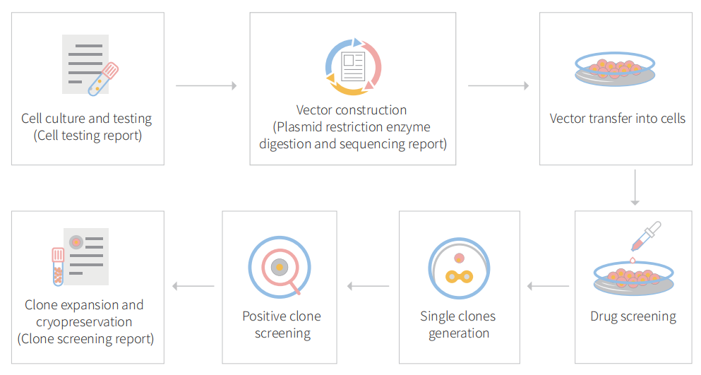
CRISPR-U™ customized workflow for engineered MDA-MB-231 model cells

Figure: CRISPR-U™ customized workflow for engineered MDA-MB-231 model cells

 Case 1: Knockout

CRISPR genome editing in triple-negative breast tumors with nanolipogel

Triple-negative breast cancer (TNBC), which has the highest mortality rate of all breast cancer, is in urgent need of a therapeutic that hinders the spread and growth of cancer cells. CRISPR genome editing holds the promise of a potential cure for many genetic diseases, including TNBC; however, its clinical translation is being challenged by the lack of safe and effective nonviral delivery systems for in vivo therapeutic genome editing. In this study, the researcher reported the synthesis and application of a noncationic, deformable, and tumor-targeted nanolipogel system (tNLG) for CRISPR genome editing in TNBC tumors. The schematic illustration of tNLG structure and in vivo CRISPR genome editing mechanism is shown in Fig1.
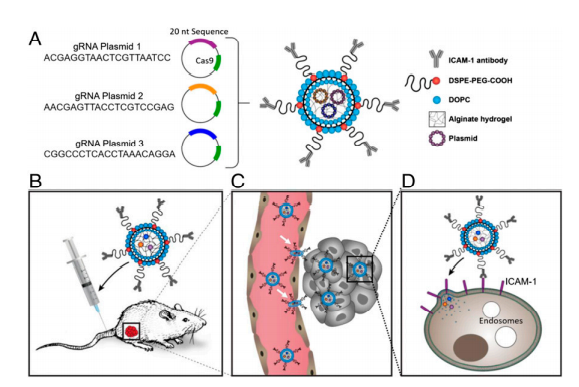

Schematic illustration of tNLG structure and bio mechanisms of in vivo CRISPR genome editing.

Fig 1: Schematic illustration of tNLG structure and bio mechanisms of in vivo CRISPR genome editing.

The researcher developed engineered tNLG as a CRISPR delivery nanovector verified its applicability in various dimensions like uniformity, core-shell nanostructure, encapsulation efficiencies, storage stability, cytotoxicity (Fig.2a-e). The in vitro TNBC specificity of tNLG in comparison with nNLG, the transendothelial capability of tNLG and tPSNP across the tumor endothelial cell (EC) barrier, and the normal EC barrier (Fig.2f,g). Deformable permeability indicated that tNLG can directly release payload into the cytosol without endosomal entrapment. Fluorescent imaging confirmed that CRISPR plasmids were delivered into the cytosol of TNBC cells by tNLGs. The cell distribution area of internalized tNLGs, indicating that internalized CRISPR plasmids were not confined within endosomes. Plasmids were successfully delivered into the nuclei of MDA-MB-231 cells by engineered tNLG (Fig. 2h-k).
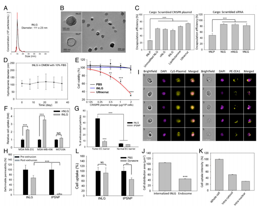

 Engineered tNLG as a CRISPR delivery nano-vector

Fig 2: Engineered tNLG as a CRISPR delivery nano-vector.

To demonstrate the therapeutic benefit of CRISPR genome editing, they selected Lcn2 as the therapeutic target for proof-of-principle TNBC-specific genome editing experiments in vitro and in vivo. Lcn2 gene expression was significantly up-regulated in human TNBC cell lines (MDA-MB-231 and MDA-MB-436) in comparison with nonneoplastic MCF10A cells (Fig. 3a,b). TNBC patients with high Lcn2 expression (cohort of 102 patients) demonstrated a significantly worse prognosis than the low Lcn2 group (cohort of 76 patients, P = 0.016; log-rank test). In vitro genome editing efficiency of tNLGs was measured by using qRT-PCR and IF staining. They found that Lcn2 CRISPR knockout in 2 TNBC cell lines did not alter their proliferation. Also, the Lcn2 CRISPR knockout did potently impede cell migration in both MDA-MB-231 and MDA-MB-436 cells (Fig.3c-l).
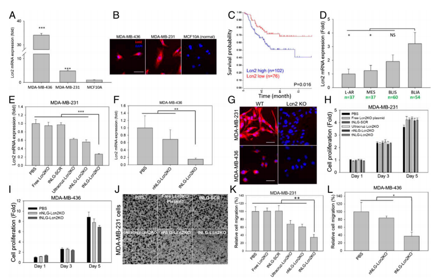

Potent in vitro CRISPR genome editing by tNLG

Fig 3: Potent in vitro CRISPR genome editing by tNLG

To evaluate the EMT phenotypical changes caused by Lcn2 CRISPR knockout, the research utilized a state-of-the-art quantitative phase imaging (QPI) method to characterize and compare a panel of cell morphological and behavioral parameters between wild-type (WT) and Lcn2 CRISPR knockout (Lcn2 KO, tNLG-Lcn2KO group) MDA-MB-231 cells. The cell motion trajectories of WT and Lcn2 KO cells are shown in Fig.4a. They found that WT cells had a significantly faster motility speed than Lcn2 KO cells, resulting in much longer migration distances (Fig. 4b-c). Besides, that WT cells exhibited a classic mesenchymal cell phenotype with significantly longer filopodia during cell migration. The Lcn2 KO cells significantly reduced their cell length and cell height resulting in significant inhibition of filopodia formation. Furthermore, Lcn2 KO cells significantly reduced mesenchymal biomarker expression and increased expression of epithelial biomarker (Fig.4d-h). Lcn2 CRISPR knockout in TNBC cells significantly reduces aggressiveness by inhibiting EMT, at least partially, and may lead to a potent in vivo therapeutic benefit in TNBC therapy.
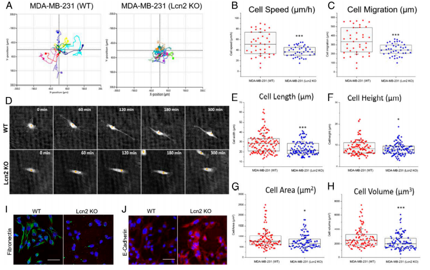

CRISPR genome editing of Lcn2 inhibits EMT of TNBC cells

Fig 4: CRISPR genome editing of Lcn2 inhibits EMT of TNBC cells

In vivo therapeutic genome editing in orthotopic TNBC tumors. The experiment was carried out by evaluating the tumor specificity and biodistribution of tNLG and nNLG in an orthotopic TNBC model using in vivo near-infrared (NIR) imaging. The therapeutic efficacy of in vivo CRISPR genome editing using an orthotopic TNBC model (Fig. 5a). Tumor progression was monitored by tumor volume measurement, (Fig. 5b). Tumor mass at endpoint (day 84) was quantified in weight (Fig.5c). Mouse body weights remained unchanged during treatment in all tested groups (Fig. 5e). They quantified the in vivo CRISPR genome editing efficiency by measuring the loss of Lcn2 gene expression in TNBC tumors using qRT-PCR as depicted in Fig. 5f as well as liver and renal toxicities of tNLG-Lcn2KO were determined by measuring serum levels of ALT, AST, Creatinine, and BUN (Fig. 5g).
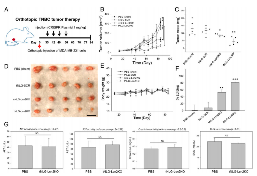

In vivo CRISPR genome editing of Lcn2 potently attenuates TNBC tumor growth

Fig 5: In vivo CRISPR genome editing of Lcn2 potently attenuates TNBC tumor growth

In summary, the researcher developed a noncationic, deformable, and TNBC-specific nanolipogel for in vivo CRISPR genome editing in human TNBC tumors. The tNLGs also represent a platform delivery system that can be used to target TNBC cells. This proof-of-principle study suggests that this tNLG formulation has a promising and broad potential for translating CRISPR genome editing into a novel precision medicine in cancer therapy.

 Case 2: Knockin

pS134-GR, a potential therapeutic target for aggressive triple-negative breast cancer

Breast cancer (BC) accounts for ~ 15% of cancer-related death in American women. Up to 40% of TNBC tumors express elevated glucocorticoid receptor (GR) levels. GR is a ligand-activated (cortisol/dexamethasone [Dex]) transcription factor member of the steroid hormone receptor (SR) superfamily and its expression associated with chemotherapy resistance and metastatic recurrence of TNBC. Although TNBC is intensely studied, molecular targeted therapies are still largely unavailable for TNBC patients, who suffer from higher disease recurrence, more frequent metastasis, and a worse prognosis. Thus, appropriate biomarkers of driver pathways and new therapeutic targets are urgently needed. Phosphorylation of GR on Serine 134 is elevated in TNBC relative to other breast cancer subtypes. ligand-dependent pS134-GR target genes are known mediators of pro-survival and metastasis in TNBC. In this study, the researcher used CRISPR/Cas9-mediated gene knockin to modify MDA-MB-231 cells to express NR3C1 containing an S134A mutation. This is accomplished using an HDR donor vector along with a CRISPR/Cas9-GFP expression vector; an anti-sense gRNA sequence (NR3C1-S134A-AS1-sgRNA) targeting the coding sequence of NR3C1 was cloned into a CRISPR/Cas9-GFP expression vector (PX458).
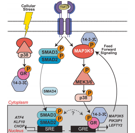

GR Ser134 phosphorylation creates a feedforward signaling loop that potentiates further activation of the p38 MAPK pathway downstream of TGFβ1 in TNBC models.

Fig : GR Ser134 phosphorylation creates a feedforward signaling loop that potentiates further activation of the p38 MAPK pathway downstream of TGFβ1 in TNBC models.

The investigator tested Dex's role in breast cancer cell migration (Fig.1). MDA-MB-231 cells were treated with increasing doses of Dex at different time points and Dex induces cellular migration was measured (Fig1. b,c). IPA analysis of MDA-MB-231 cells treated with 100 nM Dex and their pathway analysis including the TGFβ1 and p38 MAPK pathways (Fig1. d). The fraction of wound area closure of MDA-MB-231 cells treated (18 h) with vehicle control (Fig1. e).
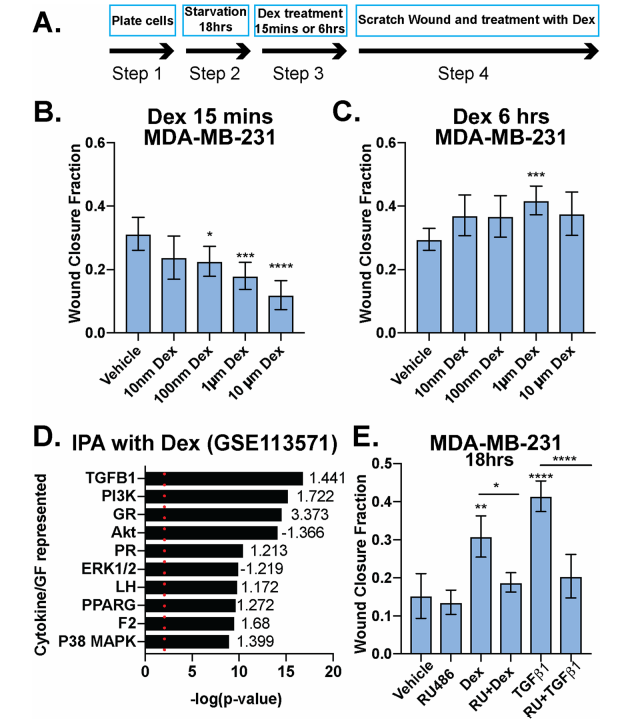

 Dexamethasone either inhibits or promotes breast cancer cell migration in a time-dependent manner

Fig 1: Dexamethasone either inhibits or promotes breast cancer cell migration in a time-dependent manner

The researcher studied the linkage between TGFβ1 signaling and phosphorylation of GR Ser134 in MDA-MB-231 cells and they found that TGFβ1 induces p38 MAPK-dependent phosphorylation of GR Ser134 shown in Fig 2a,b, and c. A Patient-derive xenograft (PDX) HCI-10 cell line was treated with TGFβ1 and evaluated the expression of pS134-GR, total GR, and total p38 (Fig.2d). Western blot analysis of pS134-GR, total GR, p-p38 MAPK, and total p38 MAPK protein levels in MDA-MB-231 cells treated with either vehicle control or HGF shown in Fig. 2e.
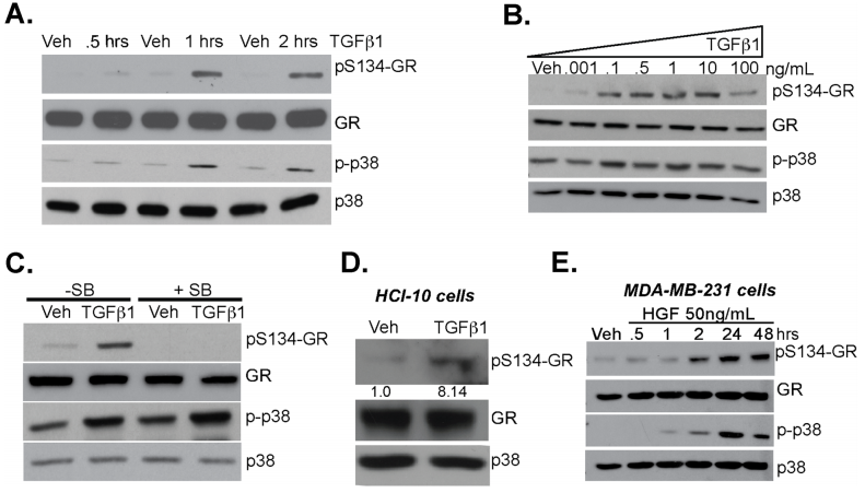

TGFβ1 induces p38 MAPK-dependent phosphorylation of GR Ser134

Fig 2: TGFβ1 induces p38 MAPK-dependent phosphorylation of GR Ser134

To test the requirement for GR Ser134 in TGFβ1- regulated TNBC cell migration, they employed a CRISPR/ Cas9 approach to creates MDA-MB-231 cells expressing either wt-GR (control) or a point mutant GR in which Ser134 has been changed to Alanine (S134A-GR clone #1 and clone #2). They assessed MDA-MB-231 cells and their clone for phosphorylation, MTT, wound healing, migratory activity, and tumorsphere assay (Fig.3 a-f). Impaired cell migration measured by chemotaxis in transwell migration assays (Fig. 3g), as well as attenuated tumorsphere formation, were also observed using the previously described GRnull/low U2OS osteosarcoma cell line engineered to stably express S134A-GR relative to wt-GR (Fig. 3h).
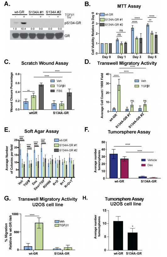

 GR Ser134 is required for TGFβ1-mediated migration of TNBC cells

Fig 3: GR Ser134 is required for TGFβ1-mediated migration of TNBC cells

The expression of 14-3-3ζ mRNA in patients with ER−/HER2- (TNBC) or Her2+ breast cancer was higher when compared to patients with ER+/HER2- breast cancer (Fig. 4a) and higher expression correlates with poor survival (Fig.4b). Besides, they observed elevated expression of 14-3-3 in different breast cancer cell lines including MDAMB-231, Hs578T, and MDA-MB-468 (TNBC) cells (Fig.4 c). They tested the necessity for 14-3-3ζ in TGFβ1-mediated migration and found that TGFβ1 induced robust cell migration in sh control but not sh14-3-3ζ TNBC models. Western blot analysis was employed to evaluate the interaction of 14-3-3ζ with GR (Fig.4d,e).
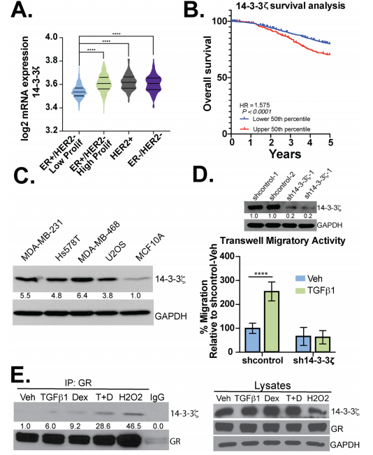

14-3-3ζ is required for TGFβ1-induced TNBC cell migration

Fig 4: 14-3-3ζ is required for TGFβ1-induced TNBC cell migration

The impact of pS134-GR on TNBC cell behavior was performed by RNA-seq studies in MDA-MB231 cells expressing either wt-GR or S134A-GR (clone #1). RNA-seq data shown the differences in transcriptomes between MDA-MB-231 cells expressing either WT or S134A-GR cells shown in Fig 5.
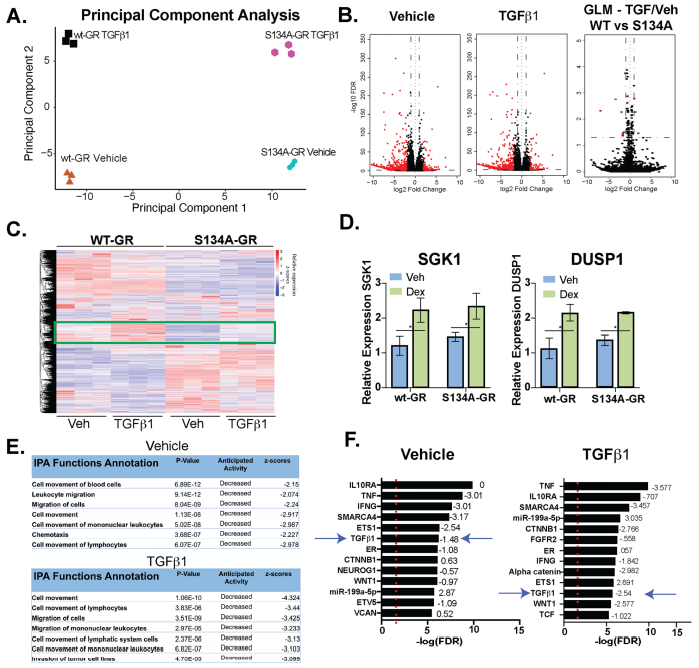

WT vs. S134A-GR transcriptomes in TGFβ1-treated MDA-MB-231 cells

Fig 5: WT vs. S134A-GR transcriptomes in TGFβ1-treated MDA-MB-231 cells

To study phosphorylation of GR Ser134 is critical for MAPK signaling, they reveal a dramatic loss of expression of genes important for MAPK signaling in TNBC cells expressing S134A-GR compared to wt-GR (Fig. 6a). Western blot analysis of different protein expression shown in knock-in, point mutation, and knockdown cell line (Fig. 6b,c,d). They also found that MAPK3K5 protein levels were significantly downregulated in two clones harboring S134A-GR relative to wt-GR (Fig.6e). MAP3K5 and GR mRNA expression levels are strongly correlated in TNBC patients (Fig.6f). Notably, inhibition of MAP3K5 with the selective inhibitor, selonsertib, blocked phosphorylation of GR (Fig. 6g) and inhibited TGFβ1-induced migration (Fig. 6h) in both MDA-MB-231 and Hs578T cells. Similarly, inhibition of p38 MAPK using SB203580, halted TGFβ1-mediated migration in both TNBC models (Fig. 6h).
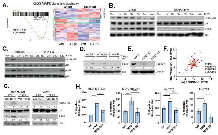

Phosphorylation of GR Ser134 is critical for MAPK signaling

Fig 6: Phosphorylation of GR Ser134 is critical for MAPK signaling

The researcher found that P-S134-GR promotes the expression of a 24-gene signature, these are correlated with poor prognosis in Breast cancer. Proteins upregulated in MDA-MB-231 cells expressing either wt-GR or S134A-GR shown in Fig7a. Gene Clustering of the 39 genes significantly upregulated in wtGR cells by TGFβ1 (Fig.7b). Representative genes (LEFTY2, PIK3IP1) were validated by qPCR in TGFβ1-treated cells predicted by RNA-seq data. They found that recruitment of S134A GR to these regions was significantly diminished (LEFTY2) or failed to occur (PIK3IP1) in CRISPR models expressing phosphomutant GR (Fig. 7d). To further evaluate the importance of the pS134-GR gene signature in breast cancer patients, they used the METABRIC dataset to calculate the average expression of the above-defined 24 pS134-GR-induced genes for each patient tumor and stratified patient populations. This difference was significant with a log-rank p-value of 0.0008. These findings in the METABRIC dataset were verified by using the SurvExpress tool with the TCGA breast cancer dataset and observed similar results with significant separation based on overall survival (Fig. 7e, f).
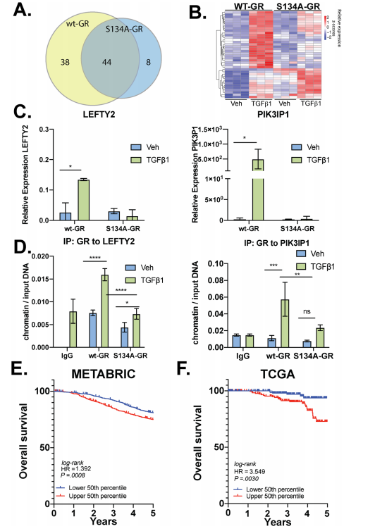

P-S134-GR promotes the expression of a 24-gene signature that correlated with poor prognosis in BC

Fig 7: P-S134-GR promotes the expression of a 24-gene signature that correlated with poor prognosis in BC

In summary, phospho-GR is a key mediator of dangerous TNBC progression. Ligand-independent but p38 MAPK-induced phosphorylation of GR on Ser134 is essential for its deleterious actions as a driver of TNBC migration, invasion, anchorage-independent cell growth, and tumorsphere formation.

Ubigene developed CRISPR-U™ which optimizes eukaryotic cells and animal gene-


editing vectors and processes. The efficiency and accuracy are 10x higher than traditional 


methods. Contact us immediately to know about your research related services!

 

Reference:

Therapeutic genome editing of triple-negative breast tumors using a noncationic and deformable nanolipogel. PNAS, 2019, 116 ( 37)18295–18303.

Glucocorticoid receptors are required effectors of TGFβ1-induced p38 MAPK signaling to advanced cancer phenotypes in triple-negative breast cancer. Breast Cancer Res. 2020, 22(1):39.

Note | The article is original by Ubigene, please indicate when reprinting.

Thursday, February 4, 2021

Trust on CRISPR to stay younger?Ubigene will tell you how likely it is!


Aging is thought of as “the time-related deterioration of the physiological functions necessary for survival and fertility”. so, it is critical for scientists to find effective ways to study the molecular drivers of aging. Although, the molecular basis of physiological aging is adequately complex that researchers have not fully unraveled the processes and genes involved in it. Simply, to date the process of aging at the molecular level is not fully explored. There are nine traits that termed as potential hallmark of aging. Genomic instability, both internal and external factors that cause genetic damage can accelerate aging. Telomere attrition, telomeres protective “caps” getting shorter each time of cell division and over time, cells loses its dividing capability, which can lead to disease. Epigenetic alterations, changes in gene expression (not changes to the DNA itself) via an individual’s life experiences or environmental factors which affect aging. Loss of proteostasis, with age, cellular proteins become misfolded and therefore, lose their homeostatic functions (damaged proteins observation with aging or age-related diseases). Deregulated nutrient-sensing, especially metabolism-regulating pathways proteins (e.g. mTOR, sirtuins) are influenced by nutrient levels and promoting aging. Mitochondrial malfunction promotes aging. Altered intercellular communication malfunction resulting in inflammation and tissue damage. Cellular senescence impaired cell cleaning process, “Older” cells deposition can lead to harmful health effects. Finally, stem cell exhaustion reduces regenerating new tissue cells sponsoring aging.

Aging is a leading risk factor for a number of debilitating conditions, including heart disease, cancer and Alzheimer's disease, to name a few. Although much remains to be discovered, patterns and connections are emerging, and this makes the needs of effective interventions for anti-aging therapies that targeting age-associated diseases. To date, researchers have found several genes in mammals that prolong lifespan, linking genetics to aging-related damage. If a gene is involved, it can be edited by CRISPR. However, editing a single gene in every cell type in the body is hard to do, if not impossible, in adults. Therefore, need to determine which cells could be edited, and which oxidative damage genes should be targeted? Similarly, instability of mitochondrial and genomic DNA are large contributors to age-related damage. This takes the form of small mutations in our DNA that occur and accumulate all over the body as cell types replenish and renew. In 2019, journal of Nature Medicine, highlight a novel CRISPR/Cas9 genome-editing therapy that can suppress the accelerated aging observed in mice with Hutchinson-Gilford progeria syndrome, a rare genetic disorder that also afflicts humans. This treatment provides important insight into the molecular pathways involved in accelerated aging, as well as how to reduce toxic proteins via gene therapy. Recent study suggests that cellular senescence is also a driver of aging and age-related pathology. Senescent cells are characterized not only by a cell-cycle arrest, but also by a potent pro-inflammatory phenotype, which is thought to drive aging and age-related diseases. Elimination of senescent cells in mice has been shown to reverse age-related pathology demonstrating that targeting senescent cells can be a powerful strategy to promote healthy aging.


CRISPR/Cas9 system can dramatically improve the physiological health and life span of human. Genome editing can provide a significant new understanding of how scientists may eventually be able to target molecular drivers of aging in humans. There will be limits to how far we can with such techniques. We could not reprogram the entire body, or enable people to live forever. But if we can repair some of the body’s worn out parts, that would allow us to enjoy a higher quality of life as we age. The newly developed CRISPR–Cas9 technology will not only support the development of genetic models of Aging & aging related diseases but also enhance our understanding of the pathogenesis of the related disorders including cancer, parkinson’s, huntington’s diseases, all of which have become more prevalent as the life expectancy of humans has increased. Novel genome editing technologies based on the CRISPR–Cas9 system together with powerful readout methods might help us better understand the logic of aging and unravel some of the mysteries of aging in general in the near future.

 Application:

CT26.WT has a wide application in the Bio-medical research arena.

1. Genetics and functional genomics: mutations affecting longevity; gene homologies; organismal and cellular aging; gene manipulation.

2. Signaling and gene expression: mechanisms linking age-associated changes with phenotypes and physiology; intracellular signaling; interactions between cells and tissues; hormonal, immune, and inflammatory systems.

3. Cell proliferation, aging and death: replicative senescence; apoptosis; telomere biology and other intrinsic and extrinsic influences; chronological cellular aging; phenotypes of aging cells.

4. Cell stress and damage: extrinsic and intrinsic influences of free radicals on cells and tissues; free radical defense and damage; free radicals as signaling molecules; stress and aging.

5. Stem cells and aging: effects of age on stem cell generation; migration and homeostasis; stem cell-niche interactions; regulatory mechanisms.

6. Integrative physiology: outcomes of aging processes at cellular, molecular and organismal levels.

7. Potential clinical applications: leading to improved prevention, treatment of disease, a greater understanding of pathological processes with aging.

8. Drugs & Aging: drug discovery, screening, optimum drug therapy.

  CRISPR-U™ gene editing for Aging research

According to WHO, at the biological level, aging results from the impact of the accumulation of a wide variety of molecular and cellular damage over time. There are nine traits that termed as potential hallmark of aging like genomic instability, telomere attrition, epigenetic alterations and loss of proteostasis etc. To date, aging researcher discovered several genes in mammals that prolong lifespan and linking genetics to aging-related damage. CRISPR-Cas system is a revolutionary genome-editing tool, which can use for precise genome editing and can generate gene knockout or knock-in genome manipulations through substitution of a target genetic sequence with a desired donor sequence. Gene-editing tools are capable of correcting gene-mediated age-related pathology, and therefore ameliorating or eliminating disease symptoms. Deleting target genes using the CRISPR system or correcting for gene mutations may ameliorate many different disorders detected in the aging population. Cancer cells targeted by the CRISPR system may result in an increased sensitivity to chemotherapeutics, lower proliferation, and higher cancer cell death. Ubigene developed CRISPR-U™ for gene manipulation of eukaryotic cell line. Thereby, possible to achieve genome editing in numerous eukaryotic cells with the utilization of the CRISPR/Cas9 system. 

 Case study :

CRISPR/Cas9 mediated study of mutation impact on telomere metabolism

Missense mutations in the human CTC1 gene cause Coats plus (CP), a rare autosomal recessive disorder characterized by retinal telangiectasia, intracranial calcifications, osteopenia, and gastrointestinal bleeding. Characterization of human disease mutations has often yielded valuable insights into basic biological functions. In this study researchers investigated the impact of CTC1L1142H on telomere metabolism and compared the effect of this CTC1 mutation in two distinct cell types, the HCT116 colon cancer cell line and the telomerase‐immortalized retinal pigment epithelial (RPE) cells. They found that mutant CTC1L1142H interacts poorly with STN1 and that the CTC1:STN1 subcomplex is sufficient to repress telomerase‐mediated telomere elongation. Expression of CP mutations that cannot interact with DNA Pol‐α show that CTC1:STN1 is also required to promote DNA Pol‐α‐mediated C‐strand maintenance. Thus, CST complex is required to coordinate both telomerasemediated Gstrand extension and DNA Polαmediated Cstrand synthesis to maintain telomere length homeostasis.

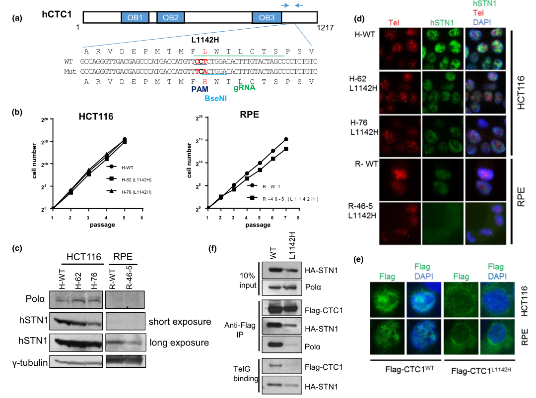
Fig 1: Generation of the CTC1 L1142H mutation in HCT116 and RPE cells using CRISPR/Cas9.


To understand mechanistically how the CTC1 L1142H mutation impacted telomere metabolism in CP patients, they utilized clustered, regularly interspaced, short palindromic repeats (CRISPR)/CRISPR-associated 9 (Cas9) to mutate CTC1 Leu 1142 to His 1142 on both alleles in the HCT116 cell line and telomerase-immortalized RPE cells. The mutant cell line were used to measure the proliferative capacities, expression pattern of endogenous DNA Pol-a and STN1,  Immuno-FISH analysis for endogenous STN1 (green) and telomeres (red) in WT or L1142H mutant HCT116 or RPE cell lines. They found that CTC1, in complex with STN1, negatively regulates telomere length. While the CTC1 L1142H mutation led to an initial increase in telomere length in RPE cells, this increase in length cannot be stably maintained.

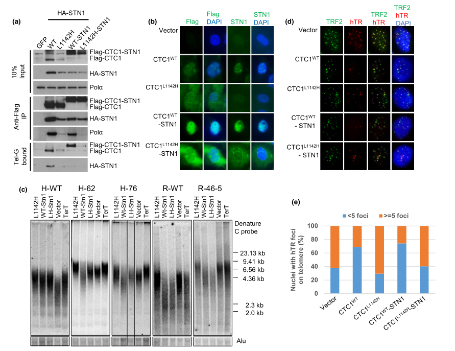
Fig 2:The CTC1:STN1 complex inhibits telomerase recruitment to telomeres.


The CTC1 L1142H mutant interacts poorly with both STN1 and DNA Pol-a and fails to bind ss telomeric DNA, suggesting that physical interactions between CTC1, STN1, and DNA Pol-a are all required to bind to ss telomeric DNA. To test this hypothesis, researcher examined whether artificially tethering mutant Flag-CTC1 L1142H to STN1 via a flexible 10-amino acid linker could rescue CTC1 L1142H ’s interaction with DNA Pol-a and ss telomeric DNA. The Flag-CTC1 WT -linker-STN1 protein interacted robustly with both DNA Pol-a and ss telomeric DNA (Figure 2a), completely localized to the nucleus (Figure 2b) and functionally reduced telomere lengths in both WT and CTC1 L1142H HCT116 and RPE cells (Figure 2c). In addition, expression of the Flag-CTC1 WT -linker-STN1 constructs reduced telomerase accumulation on telomeres, as revealed by telomerase FISH (Figure 2d and e). They found that CTC1 is required to directly interact with STN1 to form a CTC1:STN1 (C:S) complex. C:S then interacts with DNA Pol-a to enable stable binding to ss telomeric DNA, and this C:S: DNA Pol-a complex is inhibitory to telomerase-mediated G-strand extension.

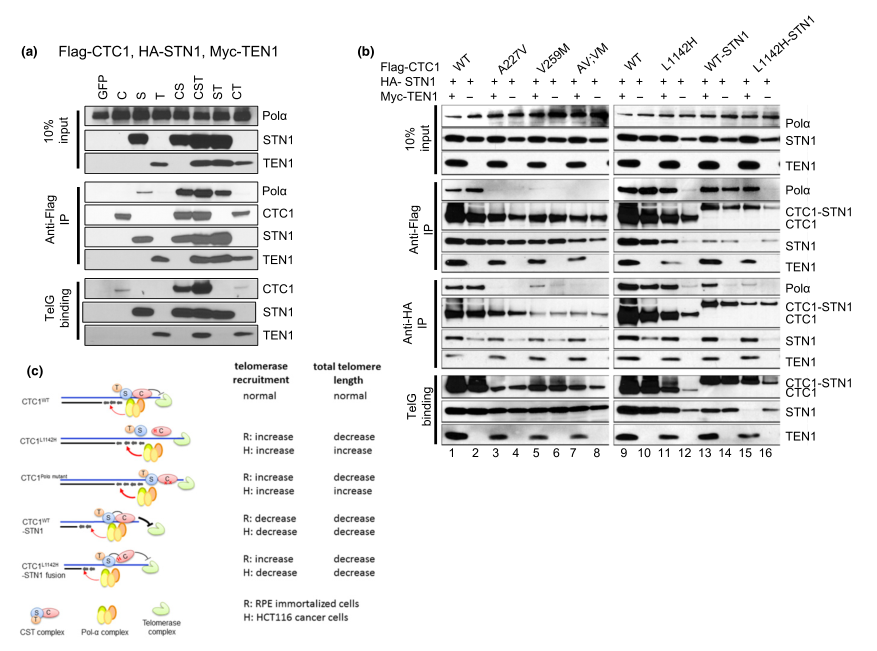
Fig 5:TEN1 enhances CTC1:STN1 interaction.


To examine the contribution of TEN1 to CST complex formation with DNA Pol-a and ss telomeric DNA, they first expressed Flag-CTC1, HA-STN1, and Myc-TEN1 in HEK293T cells and examined their interactions by Co-IP. By itself, HA-STN1, but not Flag-CTC1 or Myc-TEN1, weakly interacted with endogenous DNA Pol-a (Figure 3a). Co-expressing all three C:S:T components together resulted in robust binding to DNA Pol-a, although C:S and S:T also interacted well with DNA Pol-a. The presence of Myc-TEN1 enhanced the interaction between Flag-CTC1 L1142H and HA-STN1, as well as complex formation between Flag-CTC1 L1142H , HA-STN1, and DNA Pol-a(Figure 3b). Thus, provide functional evidence that CTC1:STN1 is required to repress telomerase activity in vivo. The CTC1 L1142H protein interacts poorly with STN1 and localizes partially to the cytoplasm, leading to telomerase-mediated telomere elongation (Figure 2c).


In summary, the CTC1 L1142H :STN1:TEN1 complex cannot compete with telomerase for access to the 3 prime G-rich overhang. Impaired interaction between CTC1 L1142H :STN1 and DNA Pol-a results in increased telomerase recruitment to telomeres and telomere elongation, further revealing that C:S binding to DNA Pol-a is required to fully repress telomerase activity. In addition, C:S regulates C-strand fill-in by DNA Pol-a. These findings confirm the CST complex as the major regulator of both G-strand extension and C-strand fill-in reactions.

Ubigene developed CRISPR-U™ which optimizes eukaryotic cells and animal gene-


editing vectors and processes. The efficiency and accuracy are 10x higher than traditional 


methods. Contact us immediately to know about your research related services!


Reference:

CTC1-STN1 coordinates G- and C-strand synthesis to regulate telomere length. Aging Cell, 2018 ;17(4):e12783.


Wednesday, February 3, 2021

H1299 cell, an ideal route for cancer mutation CRISPR therapy research|Ubigene


Lung cancer is the leading cause of cancer-related deaths worldwide, with a 5-year survival of approximately 6%. Around 80% of these are of non-small-cell (NSCLC) histological type for which surgical resection or radical chemoradiotherapy offers the best prospect of cure. Many lung cancer patients are resistant to current treatments, including chemotherapy and radiotherapy. The vast majority of cases however are diagnosed at an advanced stage, and therapy options are limited. Despite progress in research on lung cancer, targeted therapeutics, and clinical management, many knowledge gaps remain to be closed. Due to lung cancer's genetic and phenotypic diversity, individualization of therapy is becoming a reality; thus, this tumor entity is likely to further trigger future precision medicine development.

Nonsmall cell lung cancer (NSCLC) model cell line, H1299 is widely used in a variety of basic cell biology and biomedical studies involving lung cancer proliferation, corresponding inhibitors studies, tumor formation, and metastases. The researcher uses H1299 cell line as a disease model to understand the basic biology of the disease, to understand how mutations affect drug response or resistance, to understanding the mechanisms underlying differences in drug responsiveness, target identification, and validation as well as even to stratify patients for more efficient and effective clinical trials. Gene editing technologies are rapidly advancing as a realistic therapeutic option. The ability to strategically edit a patient’s genome can constitute a treatment revolution. Genome editing technologies have huge potential in lung cancer treatment including targeting oncogenes and tumor-suppressor genes, genes related to chemotherapy drug resistance, and genes related to therapies using targeted drugs and inhibitors to promote further preclinical research and the clinical treatment of lung cancer.

H1299 cell line (NCI-H1299 or CRL-5803) was established from the lung cells of a 43-year-old Caucasian male patient with non-small cell lung cancer and is widely used in biomedical research. An immortalized cell line, H1299 can divide indefinitely and the unique feature of this cell line is the lack of expression of the P53 protein, which is accounts for their proliferative propensity. This cell line (H1299) has been reported to secrete the peptide hormone neuromedin B(NMB), but not gastrin-releasing peptide (GRP) and are useful for studying lung cancer in humans. An earlier study reported that the H1299 cell line used to study genes related to sensitization and drug resistance to lung cancer chemotherapeutics such as paclitaxel, providing a theoretical basis for improving the therapeutic efficacy of chemotherapy in lung cancer. 

 Application:

Below are some applications of H1299:

1.Targeted therapy: Anti-angiogenetic drugs, Drugs targeting cell signaling, biologic mechanism, toxicity profile, Other biologic agents (vandetanib, sorafenib, etc.).

2. Drug developments: PI3K inhibitors, MEK inhibitors, EGFR inhibitors, etc.

3.  Molecular genetics: Exploitation of the genomic aberrations, somatic genetic alterations, oncogenic activation of particular tyrosine kinases.

4.  Epigenetics: Mutations in epigenetic regulators, epigenetic therapy, etc.

CRISPR-U™ gene editing in H1299 cell line

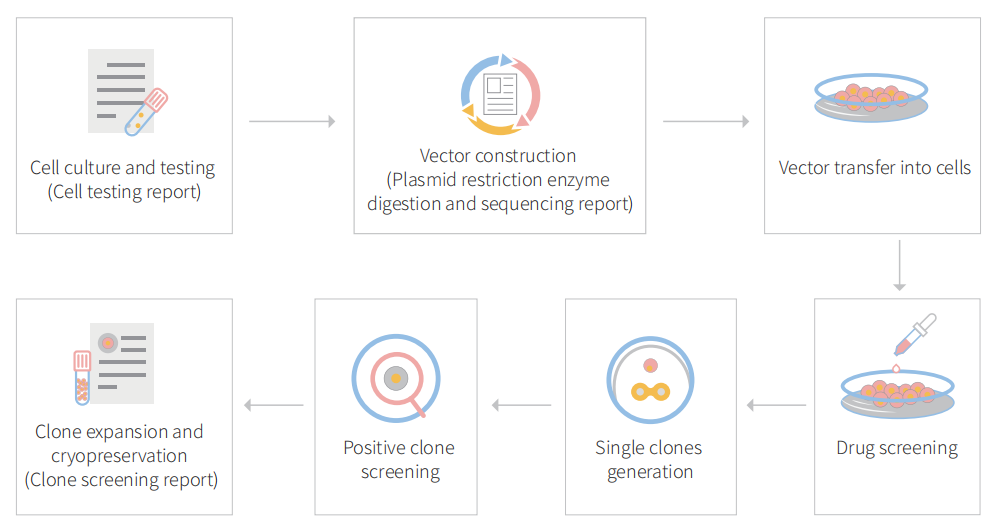
Genome editing and the creation of cellular models can advance research programs in the area of functional genomics, signaling pathways, metabolism, cell death, drug discovery, drug response, and cancer research, etc. The CRISPR system is a precise genome editing technique, which can create gene knockout or knock-in genome manipulations through the substitution of a target genetic sequence with a desired donor sequence. H1299  is a model human lung adenocarcinoma cell line, used for lung cancer study mainly drug discovery, disease mechanisms, initiation, progression, and therapeutics. CRISPR-system mediated edited H1299 cell lines allow investigators to study cancer hallmarks, disclosure of drug resistance mechanism, cancer therapeutics, cell death research, functional genomics, signaling pathways, drug discovery, drug response, and cell therapy. CRISPR/Cas9 technology positively fuel the advancement of in vivo and in vitro gene editing in lung cancer and have an immense impact on molecular medicine. Besides, knockout of the RSF1 gene in combination with paclitaxel resulted in the cell-cycle arrest in G1, increased apoptosis, and reduced cell migration and proliferation. Also, Knocking out NESTIN in H1299 cells can promote apoptosis, inhibit proliferation and colony formation, and suppress cell invasion by inhibiting epithelial-to-mesenchymal transition (EMT). Ubigene developed CRISPR-U™ for gene manipulation of the H1299 cell line. Thereby, possible to achieve genome-edited cells with the utilization of the CRISPR/Cas9 system. Ubigene can customize the gene-editing in eukaryotic cells as well as can generate various genes modification in animal models.

Figure: CRISPR-U™ customized workflow for engineered H1299 model cells

 Case study :

BCAR1 knockout cell line reveals that BCAR1 promotes proliferation and cell growth in lung adenocarcinoma via upregulation of POLR2A

Breast cancer antiestrogen resistance protein 1 (BCAR1; Crk‐associated substrate, CAS; p130cas) directly interact with the multiple protein motifs of various phosphatases and kinases, and to mediate Src through FAK bridge indirect association. BCAR1 has been reported to enhance tumor proliferation, invasion, and metastasis in several cancers (prostate cancer, endometrial adenocarcinoma, oral squamous cell carcinoma, breast cancer, and lung cancer). it also predicts poorer prognosis in lung adenocarcinoma cases. 


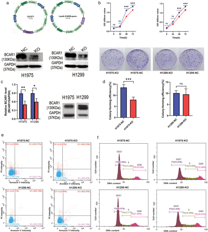
In this study, the researcher explores the role of BCAR1 in proliferation and cell growth in lung adenocarcinoma and the networks of proteins that interact with BCAR1 to trigger the proliferation of lung cancer cells. They used to lung adenocarcinoma cell line (NCl-H1975 and NCl-H1299) and established BCAR1 knockout cell line by CRISPR/Cas9 system as well as performed KO cell line for cell proliferation, colony formation, apoptosis, and cell cycle assay (Fig.1) They found that BCAR1 expression in the KO group was significantly lower than that of control as well as proliferation was significantly inhibited (Fig.1 b,c). Colony‐formation efficiency of H1975 cells was significantly decreased but no significant difference between BCAR‐KO and NC in H1299 cells (Fig.1d). Apoptosis or cell cycle progression analysis revealed that no difference in apoptosis and cell cycle of H1975 and H1299 cells following BCARKO (Fig.1 e,f). 

Figure 1: Cell proliferation, colony formation, apoptosis, and cell cycles of H1975 and H1299 cell after BCAR1 knockout


They overexpressed BCAR1 in 293T cell lines and performed immune precipitation-mass spectrometric (IP-MS) as well as different downstream analysis. The IP-MS and bioinformatic analysis predict potential interactions with BCAR1 (Fig. 2). Bioinformatic analysis revealed potential BCAR1 interact proteins and interaction partners catalytic activity and transferase activity (Fig.2a,b). The Cancer Genome Atlas (TCGA) database verification confirms BCAR1 overexpression correlates with cancer (Fig.2c). They analyze cancer-related interacting partner PPI analysis and found that high expression of POLR2A, MAPK3, MOV10, and XAB2 predicted poor prognosis in lung adenocarcinoma (Fig.2d). They consider POLR2A for further verification due to the link with more genes and the possibility of involvement in the signaling cascade.


Figure-2: Bioinformatic analysis of BCAR1 overexpressed in 293cell line


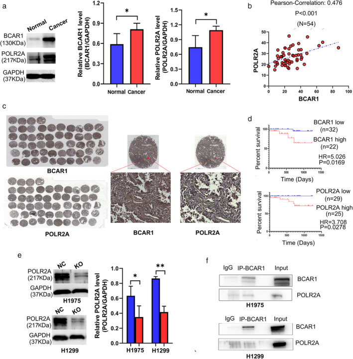
POLR2A and BCAR1 were significantly increased in lung adenocarcinoma tissues compared to adjacent normal tissues (Fig.3a). IHC‐stained TMA is shown in Fig 3b, demonstrate that BCAR1 was expressed in the nucleus, in the cytoplasm, or both locations (Fig.3c). However, POLR2A was highly expressed in the nucleus. POLR2A expression was significantly positively correlated to BCAR1 expression (R = 0.476, P < 0.001). Neither BCAR1 nor POLR2A expression was correlated with tumor size. High expression of either BCAR1 or POLR2A predicted poor prognosis in 54 lung cancer cases in the early-stage (Fig. 3d). They further verified POLR2A in H1975, and H1299 cell KO clone and western blot analysis demonstrated that POLR2A was significantly decreased in BCAR1‐KO compared to NC cells (Fig.3e). BCAR1 regulates POLR2A in H1975 and H1299 cells, despite the negative results of the CO‐IP (Fig.3f).


Figure-3: The relationship between BCAR1 and POLR2A as well as their prognostic significance in lung adenocarcinoma.


In summary, the researcher did not find a direct interaction between BCAR1 and POLR2A in H1975 and H1299 cells. The underlying mechanism of how BCAR1 and POLR2A are connected remains unclear. Moreover, synergism analysis is needed to demonstrate the role of interaction between BCAR1 and its partners concerning proliferation and cell growth in lung cancer. Future robust studies are required to resolve the interesting abovementioned points. 


[Research highlight] Enhancing p53 pathway can efficiently suppress colon cancer

  Colorectal cancer is the third most diagnosed cancer and leads to the second mortality among cancers worldwide. The first-line chemotherap...