Wednesday, January 26, 2022

[Research highlight] New target for NAFLD treatment--Metformin activates TFEB to induce autophagy


Nonalcoholic fatty liver disease (NAFLD) is a major liver disease subtype , which is characterized by an excessive hepatic lipid accumulation, covers a wide range of liver disorders, including steatosis, steatohepatitis, fibrosis, and cirrhosis. Metformin is commonly used to treat type 2 diabetes mellitus and, in recent years, it was found to play a potential role in the amelioration of NAFLD. Transcription factor EB (TFEB) is a master transcriptional regulator of lysosomal biogenesis and autophagy and, when activated, is effective against disorders of lipid metabolism.

TFEB plays a key role in lipid metabolism. However, whether TFEB is involved in the protective effect of metformin against NAFLD has not yet been reported. In response to this question, Dan Zhan et al. from first Affiliated Hospital of Kunming Medical University published an article Metformin Alleviates Hepatic Steatosis and Insulin Resistance in a Mouse Model of High-Fat DietInduced Nonalcoholic Fatty Liver Disease by Promoting Transcription Factor EB-Dependent Autophagy” in Frontiers in Pharmacology.

In this report, mice were divided into five groups: 1) the control group, 2) high fat diet (HFD)-fed group, 3) HFD + Metformin (Met) group, 4) HFD + Met + Scramble control group, and 5) HFD + Met + TFEB shRNA group. The result demonstrates that the activity of TFEB is reduced in the liver of mice fed a high-fat diet. Metformin treatment significantly reverses the activity of TFEB, and the protective effect of metformin against hepatic steatosis and insulin resistance is dependent on TFEB. They found that metformin-induced autophagy is regulated by TFEB, and the findings reveal that TFEB acts as a mediator, linking metformin with autophagy to reverse NAFLD, and highlight that TFEB may be a promising molecular target for the treatment of NAFLD. 


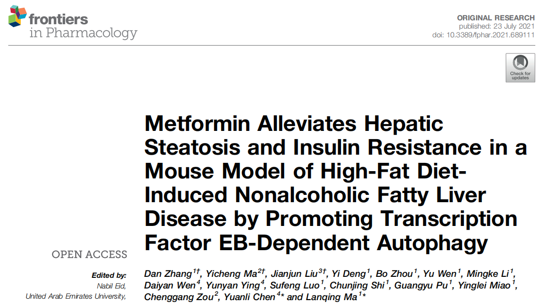
Metformin Induces Autophagy in the Liver of NAFLD Mice

A metformin treatment can signifificantly improve an excessive lipid accumulation, hepatic steatosis, and IR in the mice fed an HFD. And an autophagy dysregulation may contribute to the pathogenesis of NAFLD. So the researchers hypothesized that metformin reduces obesity, hepatic steatosis, and IR, probably via the activation of autophagy. To verify the activation of autophagy in the liver, they evaluated the levels of autophagy proteins Atg7, LC3B-II, as well as autophagic substrate p62/SQSTM1 by using Western blotting. The results demonstrate that metformin promotes autophagy in the liver of HFD-induced NAFLD mice (Figure 1).
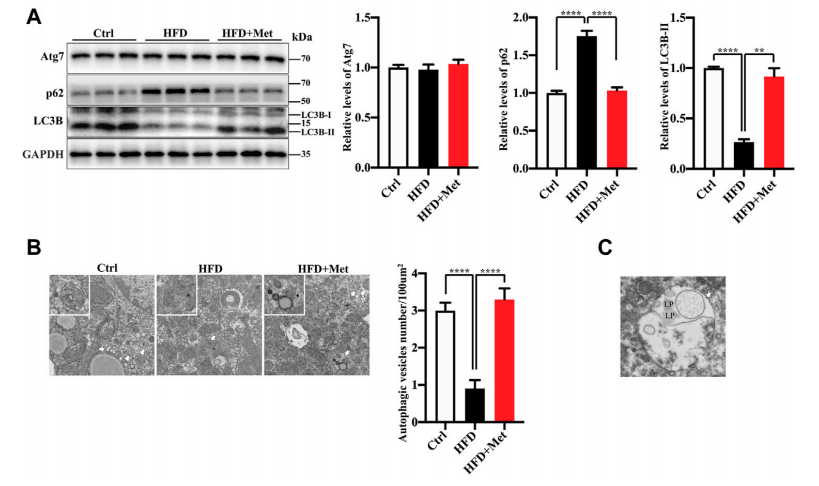


Figure 1


Metformin Induces Autophagy Through the Activation of TFEB in the Liver of NAFLD Mice

TFEB is a major regulator of autophagy and lysosomal biogenesis. The activity of TFEB mainly depends on its phosphorylation status and cytoplasm-nucleus shuttling , and the nuclear translocation is a hallmark of TFEB activation. Firstly, the researchers used Western blotting to examined whether metformin activated TFEB, the result showed that metformin treatment resulted in an increased nuclear accumulation of TFEB (Figure 2A) and reduced phosphorylation of TEFB. Then, to further test whether TFEB is involved in metformininduced autophagy, the researchers performed the knockdown of TFEB via tail vein injection with an AAV8 virus expressing an shRNA (provided by Ubigene) that targets TFEB (shTFEB). Compared with the injections of a scrambled control shRNA, the injections of recombinant AAV8-shRNA-TFEB reduced the expression of TFEB, as well as that of its target genes. Western blotting revealed that the knockdown of TFEB downregulated the expression of LC3B-II and upregulated the protein expression of p62/SQSTM1 (Figure 2C). Likewise, a downregulated TFEB level did not affect the protein level of Atg7. Taken together, these results confirm that metformin induces autophagy through the activation of TFEB.

In this study, Ubigene provided the recombinant AAV8 shRNA TFEB and the control AAV to study whether TFEB was involved in metformin induced autophagy. The AAV particles provided by Ubigene are ultra-purified, and the virus titer is 10^12~10^13 GC/ml, so it is highly adaptable to in vivo experimentsClick here to learn about our AAV production > >
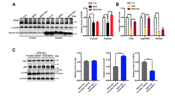


Figure 2


Metformin improves excessive lipid accumulation and IR in HFD-fed mice via TFEB

To test whether the activation of autophagy mediated by TFEB was involved in the alleviation of an excessive lipid accumulation and IR in HFD-fed mice, they measured the hepatic steatosis level using H&E and Oil red O staining after the knockdown of TFEB(Fig.3)The result showed that the protective effects of metformin on excessive lipid accumulation and IR are critically dependent on the TFEB in HFD-fed induced NAFLD mice.
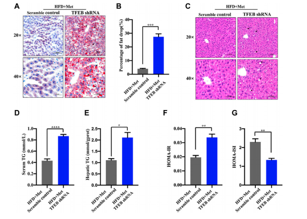


Figure 3


Overall, this study reveals a novel molecular mechanism for improving the NAFLD by using metformin, and highlight the potential beneficial effects of TFEB for the treatment of NAFLD.


Ubigene is an enterprise focusing on cell engineering. We provide high-quality gene-edited cell lines, stable cell lines, virus packaging and other related services around the world. Recentlu, Ubigene launched a KO cell bank with nearly 2000 KO cell lines. Welcome to contact our expert team to discuss your potential projects! 


More article you may interested:

How U-87 MG facilitates brain tumor research?

Study the Nobel Prize with Nobel Prize: CRISPR mediated humanized MC38 cell line


Monday, January 17, 2022

[Research highlight]A new target for human endometrial cancer treatment |Ubigene


Human endometrial cancer (EC) is one of the malignant tumors of the female reproductive tract . With the increase of aging populations, obesity, and other metabolic diseases, as well as the delay of marriage and the increasing number of infertile patients, the incidence of EC is increasing and shows a younger trend. Early diagnosis and treatment can improve the prognosis of patients with EC and reduce their clinical mortality. However, for some early asymptomatic people, the diagnosis is in the late stage of EC and the etiology of EC has not been fully clarified. Therefore, it is still an urgent issue to elucidate the mechanisms contributing to EC occurrence. 


In response to this question, Xingmei Wu et al from Lishui People's Hospital of Zhejiang province published an article “RNA Binding Protein RNPC1 Suppresses the Stemness of Human Endometrial Cancer Cells via Stabilizing MST1/2 mRNA” on MEDICAL SCIENCE MONITOR. RNA binding protein RNPC1 has been shown to play suppressive roles in various tumors, RNPC1 attenuates non-small cell lung cancer (NSCLC) progression by enhancing CASC mRNA through suppressing miR-181a binding to CASC mRNA. However, its roles in EC progression have never been demonstrated. This research shows that RNPC1 overexpression activated the Hippo pathway, a tumor-suppressive signaling, through directly binding to MST1/2 and inhibition of MST1/2 rescued RNPC1-mediated effects on EC sphere stemness.


RNPC1 was up or down regulated by overexpression lentivirus, knockdown lentivirus. All lentiviruses in this article were constructed by Ubigene (Guangzhou, China). Western blot, quantitative polymerase chain reaction and sphere forming analysis were performed to evaluate the stem-like traits of cells and RNPC1-induced effects on EC cell stemness. RNA immunoprecipitation (RIP) was constructed to investigate the underlying mechanisms. It was found that the spheres formed by EC cells, named EC spheres, exhibited a remarkably higher stemness than the parental cells, which is characterized as the increase of sphere forming ability, ALDH1 activity, stemness marker expression and migration ability. Notably, RNPC1 expression was decreased in poorly differentiated EC cells than that in EC cells with moderately differentiated. Additionally, RNPC1 expression was significantly decreased in EC spheres and RNPC1 overexpression attenuated the stemness of EC spheres. Moreover, RNPC1 overexpression decreased the migration ability of EC spheres. 

Further study showed that RNPC1 was identified as the upstream regulator of MST1/2, indicateing a possibility that targeting RNPC1 might be a good strategy to activating the Hippo pathway during EC progression, which means RNPC1 could be a potential target for EC treatment.

In this study, Ubigene provided the RNPC1 overexpressed lentivirus, knockdown (shRNA) lentivirus and empty vector to study the mechanism of inhibition of EC cell stemness by RNPC1 and MST1/2 mRNA binding. Ubigene has a mature virus packaging system and hundreds of cell transfection experience. We provide high titer viruses for in vitro cell infection and in vivo use. Now it is only $599visit us to learn more>>


RNPC1 directly bound to MST1/2 and enhanced their mRNA stability

The Hippo pathway, a tumor-suppressive signaling, the main members of this pathway are MST1/2 kinase, LATS1/2 kinase, and their junction proteins Sav and MOBI1 in mammals. The main function of the Hippo signaling pathway is to regulate the activity of transcriptional co-activators YAP and TAZ. When the Hippo signaling pathway is activated, MST1/2 kinase binds to and activates its ligand protein Sav. Then phosphorylated LATS1/2 kinase and MOB1 protein increase the formation of LATS/MOB1 complex and activate LATS1/2 kinase. Activated LATS1/2 kinase inactivates YAP and its byproduct TAZ. As the RNPC1 always binds to mRNAs and enhances their stability, the researchers wondered whether RNPC1 could activate the Hippo pathway. It was found that the expression of MST1/2 and LATS1/2 was decreased in EC spheres compared with EC cells (Figure 1A,1B). However, RNPC1 overexpression increased MST1/2 but not LAST1/2 mRNA levels in EC spheres (Figure 1C). Thus, we speculated that RNPC1 could directly bind to MST1/2 mRNA and thus activate the Hippo pathway. As expected, RIP analysis showed that MST1/2 was enriched in RNA pulled down by anti-RNPC1 but not the control IgG in EC spheres, but LATS1/2 was not enriched (Figure 1D). Additionally, MST1/2 mRNA stability was enhanced by RNPC1 overexpression in EC spheres (Figure 1E, 1F).

Figure 1


Inhibition of MST1/2 rescued the inhibition of RNPC1 overexpression on the stemness of EC spheres

Finally, the researchers explored whether RNPC1 attenuated the stemness of EC spheres dependent on MST1/2. XMU-MP-1, an inhibitor of MST1/2, was added to EC spheres with RNPC1 overexpression. The expression of the downstream effectors of MST1/2, phosphorylated LATS1/2 (p-LATS1/2), was determined to evaluate the inhibitory efficiency of XMU-MP-1 (Figure 2A). The results showed that XMU-MP-1 partially reversed the decreased ALDH1 activity (Figure 2B), the reduced sphere forming ability (Figure 2C, 2D),reduction of stemness marker expression (Figure 2E, 2F), which were mediated by RNPC1 overexpression. These results suggest that RNPC1 attenuates the stemness of EC spheres through binding to MST1/2 and thus activating the Hippo pathway.

Figure 2


In this study, RNPC1 was identified as the upstream regulator of MST1/2. However, the detailed binding regions or sites were not elucidated in this study, and therefore should be investigated in future work. Although the main conclusion should be confirmed by further in vivo experiments, targeting RNPC1 might be a good strategy to activating the Hippo pathway during EC progression.


Ubigene is a company focusing on the field of cell gene-editing. We can provide high-quality services such as virus packaging and other related services of gene editing cell line stabilization, as well as nearly 2000 kinds of KO cells in-stockGene-knockout kits and other gene-editing related products worldwide. For more details, please contact us


More articles you may interested in:

Gold standard of biological production -- CHO cell line

Versatile use of Luciferase cell line


Wednesday, January 12, 2022

U-87 MG with CRISPR/Cas9 technology can facilitates brain tumor research!


Background

In 1996, U-87 MG cell line was isolated from the malignant glioma of a 44-year-old female patient. This human primary glioblastoma cell line has epithelial morphology and can produce malignant tumors consistent with glioblastoma in nude mice. It is the most common brain tumor research model. In addition to being commonly used in brain cancer research, U-87 MG cell line is also widely used in biomedical industry, such as pathophysiology research, antibiotic screening, cancer drug research, etc. Ubigene provides wild-type U-87 MG cell line with low passage and good viability. STR authentication report is also provided. It is suitable for the construction of various gene-editing cell models.


Applications of CRISPR/Cas9 technology in U-87 MG cell line

CRISPR/Cas9 technology, as a popular gene-editing technology at present, can be used for the precise construction of various disease models, so as to facilitate research of cancer treatment, pathologyphysiology and high-throughput drug screening. This technology has been well applied to U-87 MG cell line.


Case Study 1: Gene knockout U-87 MG help to study on growth inhibition of brain tumor cells

Glioblastoma multiforme (GBM), take up 70% of all gliomas, is the most common brain tumor in adults, but there is no curative treatment at present. All patients will basically relapse even after the maximum surgical resection, radiotherapy and chemotherapy. And the median survival time is only about 15 months. Therefore, we should deeply understand the molecular mechanism of glioma occurrence and development. And looking for new targets of treatment has become one of the research hotspots in recent years.

Some studies have found that the deletion of ACSVL3 in U-87 MG will reduce its tumorigenic properties and affect signal transduction through receptor tyrosine kinase. Therefore, Kolar et al knocked out ACSVL3 from U-87 MG cell line, and conducted multiple analyses to explore the effects of ACSVL3 deletion on the carcinogenicity of U-87 MG cell line and GBM cells. Proteomic analysis showed that the content of enzymes involved in ceramide synthesis was low, the level of TCA enzyme was low and the level of glycolytic enzyme was high in ACSVL3 KO U-87 MG cell line. It was found that the knockout of ACSVL3 would affect the synthesis of lipid raft and ganglioside by fluorescence microscope and thin-layer chromatography (TLC). At the same time, the antibody detecting the mitochondrial outer membrane marker Tom20 was used for immunofluorescence. It was observed that the mitochondrial morphology of ACSVL3 KO U-87 MG was different from that of wild typeIn conclusion, Kolar et al. believe that ACSVL3 contributes to the GBM cell growth and proliferation, the synthesis of signal sphingolipids, and this enzyme may contribute to the carbohydrate metabolism involved in mitochondria [1]. This conclusion has certain research significance on how to inhibit the occurrence and expansion of GBM.

KO cell line plays an important role in studying the occurrence and expansion of tumors. Ubigene exclusive KO cell bank has nearly 2000 kinds of KO cell lines in stock, covering thousands of genes and hundreds of tumor dominated diseases, as low as $1980Click here to access our KO cell bank>>


Case study 2: U-87 MG point mutant cell line contributes to the pathophysiology of Gaucher's disease (GD)

Gaucher's disease (GD) is a disease caused by acid β-Autosomal recessive lysosomal deposition caused by glucosidase gene (GBA1) mutation. Pavan et al. Used CRISPR/Cas9 to edit GBA1 in human monocyte THP-1 cell line and glioblastoma U-87 MG cell line to construct isogenic GD model (GBA1 mutant). It was found that the two edited cell lines showed low levels of mutant acidity β-Glucosidase expression, less than 1% residual activity, and a large amount of substrate accumulation. In addition, GBA1 mutant U87 MG showed that the mutant enzyme was retained in the endoplasmic reticulum and degraded by proteasome, triggering the unfolded protein reaction (UPR). Compared with wild-type cells, the accumulation of α-syn in GBA1 mutant U87 MG could increase interleukin-1β, and the mutant cell line has higher cell mortality [2]. These results provide a theoretical basis for the study of the pathophysiological mechanism of GD.

The construction of point mutation cell model is helpful to study the pathological mechanism of single gene mutation diseases, but the successful construction of the model not only needs to select appropriate gene-editing method, but also needs rich experience of cell line constructionUbigene specialized in point mutation cell disease model generation, as low as $8480, exclusive CRISPR-U, higher success rate! For more promotion details, please click here>>


Case study 3: Multiple U-87 MG point mutation cell lines contribute to cancer therapy development

Antibody drug conjugates can target cells and genes with high specificity. Kwon et al constructed multiple variants of TP53 gene in U-87 MG using CRISPR/Cas9 system, and determined that the main transcriptional difference was due to the loss of p53 function. Using transcriptome data, they predicted which mutant clones had less phenotypic difference from the wild-type, so as to screen the best candidate genes that can be used as a drug delivery test platform. Further experiments on cell morphology, proliferation rate and target antigen mediated uptake also confirmed their hypothesis. According to the results of comprehensive analysis, they had successfully screened the most suitable mutant clone. This study provides a great idea for exploring cancer therapeutic reagents [3].


Ubigene, taking the advantage of exclusive CRISPR-U™ Technology, provides high-quality and mature gene-editing cell services, including gene knockout, point mutation, knockin. In addition, our KO cell bank has nearly 2000 KO cell line in stock. Welcome to contact us for more details! 


More articles you may interested in:

4 Must-know tips of purchasing cell lines

How does CRISPR/Cas9 work for RKO cell line research?


Tuesday, January 4, 2022

[Research highlight] A new idea of prevention and cure of COVID-19! |Ubigene


SARS-CoV-2 has caused a worldwide pandemic since 2019. Some studies have found that SARS-CoV-2 increases the number of autophagosomes, which seems to be a good breakthrough in prevention and treatment. However, a study on non-human primates showed that the FAD approved potential drug candidate chloroquine (CQ)/hydrochloroquine, as an autophagy regulator, could not inhibit SARS-CoV-2 infection, and it was found that it had no beneficial effect on the mortality of COVID-19 patients.


In view of this situation, Chao Shang, etc. (Changchun Veterinary Research Institute, Chinese Academy of Agriculture Sciencesbelieved that the mechanism of SARS-CoV-2 regulating autophagy in the host needs to be fully studied in different animal models. Recently, they published in the Journal of Virology (2020 IF: 5.1027) "Inhibition of autophagy suppresses SARS-CoV-2 replication and ameliorates pneumonia in hACE2 transgenic mice and xenografted human lung tissues" Thresults of SARS-CoV-2 infection on the established non-human primate Macaca fascilaris model and human angiotensin converting enzyme 2 (hACE2) transgenic mice, and constructs the original SARS-CoV-2 infection model based on human lung xenotransplantation mice have been shown in the article. It reveals the new mechanism of SARS-CoV-2 manipulating autophagosome formation, and found that autophagy inhibitors may be potential drugs for the treatment of SARS-CoV-2 virus infection, which provides a new idea for the study of the pathogenic mechanism of SARS-CoV-2 and its prevention and treatment.


SARS-CoV-2  infection was found to trigger autophagy in the Macaca fascularis infection model

Researchers analyzed autophagy by collecting SARS-CoV-2 infection in the lungs of the Macaca fascularis and the corresponding areas of the infected lung. It was found that the expression level of Atg5-Atg12 and LC3-I /LC3-I was significantly increased after SARS-CoV-2 infection (Autophagy activation).


There are two main ways for SARS-CoV-2 to increase autophagy

LC3-I, which is mainly located in the cytoplasm, is transformed into LC3-II after autophagy activation. LC3-II is integrated into the extended phagocyte membrane and controlled to extend into a complete autophagosome. Therefore, LC3-II can be used as a reliable autophagosome marker. Western blot (WB) showed that the expression level of LC3-II in Vero E6 and Huh-7 cell lines increased significantly after SARS-CoV-2 infection. In order to visually observe the accumulation of autophagosomes, researchers constructed Vero E6 and Huh-7 cell lines transformed with GFP-LC3 plasmid (constructed by Ubigene), and found that the level of GFP in cells increased significantly after SARS-CoV-2 infection for 24 hours (hpi), indicating that SARS-CoV-2 infection induced the formation and accumulation of autophagosomes. In addition, EGFP-mCherry-LC3 transient Vero E6 cell line (constructed by Ubigene) was constructed to visualize the transfer of LC3 from autophagosome to lysosome. It was found that SARS-CoV-2 increased the number of autophagosomes in two ways: (1) the biogenic events of autophagosome increased; (2) the fusion of autophagosome and lysosome decreased.


Figure 1 SARS-CoV-2 induces autophagosome formation in cell lines and blocks autophagosome-lysosome fusion


Autophagy related cascade reactions were analyzed to determine the relationship between autophagosome formation and virus replication

In order to further clarify the mechanism of SARS-CoV-2-induced autophagosome formation, researchers analyzed a series of signal cascades involved in autophagosome formation in the Vero E6 cell line infected with SARS-CoV-2. mTORC1 signaling pathway is the most common upstream autophagy inhibitory regulator. Researchers choose to monitor its activity by phosphorylation of its downstream substrate. It was found that SARS-CoV-2 not only inhibited Akt-mTOR signaling pathway, but also activated ULK-1-Atg13 and VPS34-VPS15-Beclin1 complexes. Moreover, in order to determine the relationship between autophagosome formation and virus replication and the key factors of virus replication, researchers used broad-spectrum autophagy inhibitor 3-MA and VPS34 inhibitor SAR405. It was found that the pretreatment of 3-MA and SAR405 significantly reduced the virus load in the Vero E6 cell line, indicating the importance of autophagy formation and VPS34 complex activation for SARS-CoV-2 virus replication.


Figure 2 Autophagy formation and VPS34 complex activation promote the replication of the SARS-CoV-2 virus


Inhibition of autophagy can reduce SARS-CoV-2 replication and improve pneumonia in mice

Researchers used two different animal models to simulate the infection process of the SARS-CoV-2 in vivo. In the hACE2 transgenic mouse model, inhibition of autophagy can inhibit the replication of SARS-CoV-2 virus and improve pneumonia. On the contrary, rapamycin induced autophagy can promote viral autophagy and aggravate the pathological process of pneumonia.


In the innovative mouse model (transplanted with human lung tissue), they proved that SARS-CoV-2 could replicate itself in human lung tissue and spontaneously produce pneumonia.


Figure 3 Inhibition of autophagy can inhibit SARS-CoV-2 replication and improve pneumonia in the mouse model (transplanted with human lung tissue)


In conclusion, researchers confirmed that autophagy promotes the replication of SARS-CoV-2 and promotes the process of pneumonia, and proposed that autophagy inhibitors may be potential drugs for the treatment of SARS-CoV-2 virus infection. Through the mutual verification of in vivo and in vitro experiments. This study lays a theoretical foundation for the in-depth study of virus cell interaction in the future, and provides a useful idea for the development of autophagy related antiviral drugs.

Ubigene focuses on cell line engineering. In this study, engineered Vero E6 and Huh-7 cell lines were provided to explore the relationship between SARS-CoV-2 virus replication and autophagy induction mechanism.


Ubigene specializes in gene editing cell line, stable cell line generation,  virus packaging, etc.  Ubigene has about 2000 in-stock KO cell lines in our exclusive KO cell line bank, and EZ-editor™ Gene-editing Series Products, such as Gene Knockout kit help to achieve successful gene-editing! For more information about our services, please inquire >>

Monday, January 3, 2022

gene-editing MC38 cell line:study the Nobel Prize with Nobel Prize


Background

MC38 cell line was derived from the colon adenocarcinoma cell line of C57BL/6 mice, which is adherent growth and fibroblast morphology. MC38 cell line implanted into C57BL/6 mice or mice with the low immune function will form tumors and metastases, which are mostly used to study the occurrence and metastasis of colorectal cancer and become a common way to verify the efficacy of tumors[1]At present, the commonly used cell models are lentivirus-mediated stable cell line models and CRISPR/Cas9 mediated gene-editing models. Ubigene has been committed to stable cell line generation and gene-editing cell line generation. Ubigene has successfully modified genes from more than 100 cell lines, including the MC38 cell line. We are providing services all over the world. For more details, please inquire>>


MC38 cell line and CRISPR/Cas9 technology can assist tumor immunotherapy

As the Nobel Laureate in 2020, CRISPR/Cas9 technology has already been used in many biological and medical fields. The combination of CRISPR/Cas9 technology and the Nobel Laureate in 2018 "tumor immunotherapy" brings more hope for tumor treatment.


The key gene for validation, which is expected to find new therapeutic targets for colon cancer

Sialylated glycan structures are known for their immunomodulatory capacities and their contribution to tumor immune evasion. However, the role of aberrant sialylation in colorectal cancer and the consequences of complete tumor desialylation on antitumor immunity remain unstudied. Cornelissen et al. used CRISPR/Cas9 technology to KO CMAS gene in MC38 cell line and found that compared with control MC38 cell line, mouse colorectal cancer MC38 cell line (MC38 Sianull) with complete knockout of CMAS gene significantly promoted tumor growth in vivo (Fig. 2). The enhancement of tumor growth caused by MC38 Sianull cell line can be attributed to the decrease of CD8+T cell frequency in the tumor microenvironment. MC38 Sianull cell line can induce CD8+T cell apoptosis in an antigen-independent manner, but the frequency of immune cells in tumor-draining lymph nodes was not affected. And in human colorectal cancer cells, low CMAS gene expression is associated with reduced recurrence free survival. In conclusion, these results show that CMAS gene is one of the key genes of sialylation pathway, which provides a new research idea for the treatment of colon cancer[2].


Figure 1  Different effects of CMAS gene knockout MC 38 cell line and control MC38 cell line on tumor growth in vivo

Reveal the relationship between Tn antigen and immunosuppressive tumor microenvironment

Expression of the tumor-associated glycan Tn antigen (αGalNAc-Ser/Thr) has been correlated to poor prognosis and metastasis in multiple cancer types. However, the exact mechanisms exerted by Tn antigen to support tumor growth are still lacking. Therefore, Lenneke et al. explored how Tn antigen affects tumor immune cell formation in a colorectal cancer (CRC) mouse model. They found that C1galt1c gene knockout with CRISPR/Cas9 technology could lead to the increase of Tn antigen level on the cell surface of MC38 cell line (MC38-Tnhigh). RNA sequencing and subsequent GO term enrichment analysis of our Tnhigh glycovariant not only revealed differences in MAPK signaling and cell migration, but also in antigen processing and presentation as well as in cytotoxic T cell responses. The results show that  MC38-Tnhigh tumors displayed increased tumor growth in vivo, which was correlated with an altered tumor immune cell infiltration, characterized by reduced levels of cytotoxic CD8+T cells and enhanced accumulation of myeloid-derived suppressor cells. But there is no systemic differences in T cell subsets were observed[3]


hCD47-MC38 HuCELL can be used as a model for the in vivo evaluation of human SIRPα antibodies

CD47 is broadly expressed on all normal cell lines or tissues, SIRPα(Signal regulatory proteinα) is a typical inhibitory immune receptor in SIRP family. After binding with CD47, it can prevent cell lines from being swallowed by macrophages. Daniel Xianfei et al. developed the human CD47 and SIRPα double knock-in mouse model (CD47/SIRPα HuGEMM™) via CRISPR/Cas9 technology. And they have also generated mouse syngeneic MC38 cell line to express human CD47 (hCD47-MC38 HuCELL™). They transplanted hCD47-MC38 HuCELL to CD47/SIRPα HuGEMMHugemm mice, which was used to evaluate the efficacy of anti human CD47 antibody Hu5F9. The experiment results show that hCD47/hSIRPα double knock-in model and hCD47-MC38 HuCELL provide a powerful preclinical platform, which can not only effectively evaluate the action mode and in vivo efficacy of human specific anti-CD47 antibody, but also can be used as a model for in vivo evaluation of human SIRPα antibodies, and a proof of concept for combination therapy with other immune checkpoint inhibitors or immunomodulators[4].


Ubigene can achieve higher cutting efficiency with developed CRISPR-U™ technology. And Ubigene has successfully modified more than 5000 genes from over 100 cell lines, including the MC38 cell line. Ubigene provides services all over the world. Now Ubigene is offering a time-limited promotional price for gene-editing cell line services, including Knockout/knockin/point mutation/overexpression.  For more details, please feel free to contact us!


References

[1] Zhou, Xiu-Man, et al. "Intrinsic expression of immune checkpoint molecule TIGIT could help tumor growth in vivo by suppressing the function of NK and CD8+T-cells." Frontiers in immunology 9 (2018): 2821.

[2] Cornelissen, Lenneke AM, et al. "Disruption of sialic acid metabolism drives tumor growth by augmenting CD8+ T cell apoptosis." International journal of cancer 144.9 (2019): 2290-2302.

[3] Cornelissen, Lenneke AM, et al. "Tn antigen expression contributes to an immune suppressive microenvironment and drives tumor growth in colorectal cancer." Frontiers in oncology 10 (2020): 1622.

[4] He, Daniel Xianfei, et al. "Anti-human CD47 antibody Hu5F9 inhibits MC38 tumor growth in hCD47/hSIRPα double knock-in mice." (2020): 5615-5615.

[Research highlight] Enhancing p53 pathway can efficiently suppress colon cancer

  Colorectal cancer is the third most diagnosed cancer and leads to the second mortality among cancers worldwide. The first-line chemotherap...