Tuesday, May 19, 2020

CircRNA Editing | Ubigene

circRNAs are circular noncoding RNAs formed by reverse splicing of pre-mRNAs. circRNAs were firstly found in viruses in the 1970s. However, due to the extensive use of the method for enrichment of poly (A) (no 5 'and 3' ends of circRNA) in the early RNA library preparation, and the calculation algorithm that RNA-seq reading requires linear alignment with the genome, a large number of circRNA information was omitted, which led to the belief that circRNA is just a byproduct of miss splicing.

With the development of high-throughput sequencing technology and bioinformatics, thousands of circRNA have been found, and more and more basic researches related to circRNA have been done. A large number of studies have shown that circRNA is endogenetic, abundant, conservative, and stable in mammalian cells, and often shows tissue or space-time specificity. It can participate in the regulation of cell growth and development, as well as the occurrence and development of diseases through a variety of mechanisms. Therefore, in recent years, circRNA has become popular in the field of non-coding RNA research

According to the origin of circRNA, it can be sorted into three types:

1) Exonic circRNAs: All sequences are derived from exons.

2) EIciRNAs: Sequences are derived from exons and introns.

3) ciRNAs: All sequences are derived from introns.

circRNA is formed by the pre-mRNA by back splicing. At present, there are three kinds of mechanisms reported as follows:

1) Intron reverse complementary sequence

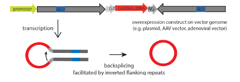
The flanking introns at both sides of the exon contain many pairs of reverse complementary sequences. The reverse complementary sequence promotes the intron sequence pairing, making the Splice-Donor in the downstream close to the Splice-Acceptor in the upstream, so as to form a circRNA. (Fig 1. Left)

2) RNA binding protein

The flanking introns at both sides of the exon contain the motifs recognized by RNA binding proteins (RBPs). RBP, when combined with the specific motifs of the two flanking introns, will form dimers, promote the two flanking introns close to each other, and then connect to form a ring. 

3) Lariat-driven circularization

When the pre-mRNA is spliced, exon skipping occurs, which results in the formation of a lariat intermediate containing exon and intron. Then the intermediate is back spliced to form a circRNA. 

The most common function of circRNA is to bind to miRNA as miRNA sponge, thus affecting the regulation of miRNA on genes.

Many circRNAs contain protein binding sites, which can be used as protein sponges.

In addition to being miRNA and protein sponge, circRNAs can also be used as a scaffold protein to promote the co-location of the enzyme, to inhibit the target gene expression by binding transcription factors, to participate in the regulation of parent gene expression, and to translate polypeptides under specific circumstances. According to the different functions, the locations of circRNAs are different. For example, as a miRNA or protein sponge, circRNA needs to be transported from the nucleus to the cell-matrix to play a role. When participating in the regulation of parent gene expression or binding transcription factor to inhibit the target gene, circRNA often plays a role in the cell nucleus.

Reference

Kristensen, L. S., Andersen, M. S., Stagsted, L. V., Ebbesen, K. K., Hansen, T. B., & Kjems, J. (2019). The biogenesis, biology and characterization of circular RNAs. Nature Reviews Genetics, 20(11), 675-691.

Relationships between circRNAs and diseases

At present, the most studied is the relationship between circRNAs and tumors. Some circRNAs promote tumor formation, such as circPvt1 in squamous cell carcinomas of the head and neck, cirs-7 (CDr1as) in colorectal cancer, esophageal squamous cell carcinoma and hepatocellular carcinoma. Some circRNAs suppress tumors, such as circsMARCA5 and circ-SHPRH in glioblastoma. Some circRNAs may play different roles in different tissues or cells, such as circHiPK3, which is a proto-oncogene in rectal cancer but suppresses cancer cells in bladder cancer.

In addition to cancers, circRNA has been found to be closely related to diabetes, cardiovascular disease, chronic inflammation and nervous system diseases. It is believed that with the development of biotechnology and more in-depth researches on circRNA, the formations and mechanisms of circRNAs can be identified. 

circRNAs can play important roles in disease prevention, diagnosis and treatment discovery.

circRNA related custom services:

circRNA involves many complex functions, so how to study its functions? Similar to protein-coding genes, the most common methods are knockout, knockdown (RNA interference) or overexpression of the circRNA. 

circRNA knockout

circRNA knockout refers to editing at the level of DNA to achieve the purpose of a complete knockout. gRNA and Cas9 would be transferred into cells by virus transduction or nucleofection. After drug screening, single clones would be generated. Positive clones would be validated by sequencing.

The most common startegies for circRNA knockout

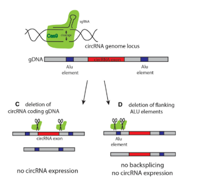
Strategy 1The most commonly used method is to design two gRNAs at both ends of the circRNA exon to knockout the whole cyclized exon sequence. Although this strategy can knockout the circRNA, it will also affect the parent gene encoding the protein, and the study on its function is not ideal.

Strategy 2The ideal method is to knockout the loop forming elements (Alu) in the flanking intron of the exon, so as to destroy the circRNA loop forming without affecting the expression of the coding gene.Fig 4.)

Ubigene is experienced in designing the strategy of a knockout the loop forming elements in the flanking intron of circRNA exon, to achieve the purpose of knockout circRNA without affecting the expression of the coding gene. Combined with CRISPR-UTM technology, the positive clones of circRNA knockout can be generated 10x faster than other common methods.

Case study

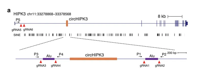
circ-HIPK3 is a kind of circRNA rich in human cells, which can combine with a variety of miRNAs as a regulator of cell growth and affect the formation of tumors. In order to verify how circ-HIPK3 forms into a circle, it is necessary to find the loop forming elements in flanking intron. A pair of sgRNA is designed for the two Alu elements predicted at upstream and downstream respectively. The predicted loop forming elements are knockout by CRISPR/Cas9 system to detect whether the expression of circRNA changes. After PCR and RT-qPCR verification, it was found that the expression of circ-HIPK3 was significantly down-regulated after knockout of the downstream loop forming elements, while the expression of circ-HIPK3 was not decreased but increased after knockout of the upstream loop forming elements. It was speculated that there were too many loop forming elements in the upstream and the prediction was not accurate. In order to further verify the RNA circulation driven by other elements, the large fragment of the intron in the upstream of the element was knockout by co-injection of gRNA3 or gRNA4 with gRNA5 or gRNA6. RT-qPCR results showed that the expression of circ-HIPK3 decreased, indicating that other loop forming elements of circ-HIPK3 exist.

Reference

Zheng, Q., Bao, C., Guo, W., Li, S., Chen, J., Chen, B., ... & Liang, L. (2016). Circular RNA profiling reveals an abundant circHIPK3 that regulates cell growth by sponging multiple miRNAs. Nature communications, 7(1), 1-13.

circRNA knockdown (RNAi)

Among the methods to study the function of circRNA, the most classical way to inhibit circRNA is to knockdown it by RNAi (shRNA or siRNA). In order to avoid affecting mRNAs of coding genes, the shRNA should be designed at the back splicing site (BSS).

Ubigene can design high-score shRNA and use lentivirus to transfer the RNA interference vector into the cells. Cells were screened according to the drug screening, and the stable cell lines with circRNA knockdown were obtained

Case study

siRNA was used to interfere with circ-HIPK3, and whether the knockdown of circ-HIPK3 would affect cell proliferation or apoptosis was observed. First, three groups of experiments targeting the linear transcript of HIPK3 mRNA, circ-HIPK3 circular transcript, and both two transcripts. The designed siRNA interfered with the corresponding transcripts was verified on the HEK-293T cell line. 

Cell proliferation and apoptosis were detected by CCK-8 and EdU assays. The results showed that the knockdown circ-HIPK3 significantly inhibited cell proliferation.

Reference

Zheng, Q., Bao, C., Guo, W., Li, S., Chen, J., Chen, B., ... & Liang, L. (2016). Circular RNA profiling reveals an abundant circHIPK3 that regulates cell growth by sponging multiple miRNAs. Nature communications, 7(1), 1-13.


Gene-Editing Liver Cancer Cell Line HepG2 - An Artifact for Research in Drug, Hepatotoxicity and Can

As one of the five internal organs in the human body, the liver is vital to the body's metabolism, detoxification, blood coagulation, and other essential processes. Participating in the body's immune system, the liver is an indispensable organ for maintaining the body's normal operation. In the study of liver diseases, one of the most useful tools is to create a suitable cell model. However, due to the difficulties in the obtainment and culturing of conventional liver cells, the development of liver disease research has been confined in some ways.  Moreover,  the relatively high cost of culturing in conventional liver cells also makes it less likely to carry out the research. Therefore, selecting liver cell lines with simple culture and stable genetic background has become an alternative in liver research, and HepG2 is one of the most commonly used liver cancer cell lines.


 The application of HepG2 cell line

HepG2 was established in 1979 by Knowles et al. It is a hepatoblastoma derived from a 15-year-old Caucasian male liver cancer specimen. HepG2 cells have an epithelial-like morphology, with a typical chromosome number of 55.

1. Hepatitis virus research: HepG2 does not contain the hepatitis B virus (HBV) or hepatitis C virus (HCV), so HepG2 is a commonly used model for studies in HBV and HCV cell lines.

2. In vitro HCC model: HepG2 is a cell line derived from liver tissue of patients with hepatocellular carcinoma (HCC) and it is a common in vitro HCC model. There is no mutation in p53 tumor suppressor gene in HepG2, so it can be used to study the close relationship between p53 and hepatocellular carcinoma (HCC), as well as the onset, diagnosis, treatment and prevention of HCC.

3. Drug research: The liver is the most essential organ for detoxification and drug metabolism in the human body. HepG2 cell line is closer to human liver tissue both in terms of morphology and functions. Therefore, this cell line is often used in drug metabolism and liver toxicity studies.


The combination of liver cell line and CRISPR / Cas9 helps to study medical problems such as hepatitis, genetic diseases and cancer

Applying CRISPR / Cas9 to construct HepG2 knock-out HBV infected cell models provides new ideas for the complete cure of hepatitis B

Hepatitis B virus (HBV) is a DNA virus. The genomic DNA of HBV outside the cell is double-stranded relaxed circular DNA (rcDNA). When the virus infects the cell, the HBV genome will enter the nucleus of the host cell. Then the rcDNA will be converted into covalently closed circular DNA (cccDNA) and exist stably in the cell. At present, an important reason for not being able to completely cure hepatitis B lies in the difficulties in clearing the stable cccDNA in the liver cell nucleus. The conversion of HBV rcDNA to cccDNA is a necessary step for the establishment of persistent infection of hepatitis B virus. Therefore, the study of the molecular mechanism of HBV cccDNA formation has far-reaching significance for the treatment of hepatitis B.

In order to explore the mechanism of HBV cccDNA formation, researchers used HepG2 to express the HBV receptor, the sodium ion-taurocholic acid co-transporter (NTCP). They established an efficient HepG2-NTCP cell model for HBV infection, which could detect the formation of cccDNA after only 24 hours. Afterward, the experiments using viral DNA polymerase inhibitors and siRNA (targeting the host DNA polymerase) showed that HBV viral polymerase was not a critical factor in the conversion of rcDNA to cccDNA. However, the knockdown of POLK, POLL, and POLH can effectively reduce the expression levels of viral nucleocapsid protein (HBeAg) and 3.5 kb RNA. Among the three genes, the knockdown of POLK showed the most significant influence in the inhibition of HBV infection.

Furthermore, using CRISPR / Cas9 to knock out POLK in HepG2-NTCP cells can greatly decrease the formation of cccDNA, confirming that the formation of cccDNA in HBV requires the participation of POLK. POLK replenishment in POLK knock-out HepG2-NTCP cells can restore HBV cccDNA synthesis and the expression of viral protein.

This study strongly indicated that POLK is a key cell molecule for HBV cccDNA formation in the HBV-infected HepG2-NTCP cell model, which provides new ideas for research in the molecular mechanism of cccDNA formation and new treatments for chronic hepatitis B.

CRISPR-U ™ can transfer CRISPR / Cas9 into Hep-G2 cells 10 times more efficiently, and select suitable cells for monoclonal culture after screening. Different clones were selected for target site amplification and sequencing verification separately, after which the positive clones with the knockout gene were selected.

Building HepG2 model of missing mutant cells by CRISPR / Cas9 revealed the causes of Wilson disease (hepatolenticular degeneration)

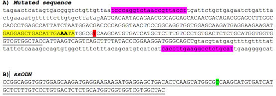
Hepatolenticular degeneration (Wilson disease) is a recessive genetic disorder caused by pathogenic loss-of-function variants in the ATP7B gene. It is characterized by disrupted copper homeostasis resulting in liver disease and/or neurological abnormalities. The variant NM_000053.3:c.1934T > G (Met645Arg) has been reported as compound heterozygous, and is highly prevalent among Wilson disease patients of Spanish descent. Accordingly, it is classified as pathogenic by leading molecular diagnostic centers. However, functional studies suggest that the amino acid change does not alter protein function, leading some researchers to question its pathogenicity. Here, we used a minigene system and gene-edited HepG2 cells to demonstrate that c.1934T > G causes ~70% skipping of exon 6.

The CRISPR / Cas9 and ssODN were co-transformed into HepG2 cells by nuclear transfection and then selected for monoclonal culture after screening. The target sites were amplified and sequenced separately for the monoclonal, and the following 4 clones were screened for research.

Compared to the transcription in wild-type cells, compound heterozygous HepG2 cells (2F3 clone) express only 15% of the transcript containing exons 5, 6 and 7. Compared to wild-type cells,  and homozygous cells (clones 1E8 and 1F6) express only 31–33%.

In addition, protein lysates were extracted from wild-type and edited HepG2 cells to determine ATP7B protein expression by western blot. Compared to wild-type cells, compound heterozygous (2F3) and homozygous (1E8, 1F6) cells express reduced levels of ATP7B, with increased ATP7B expression in 1E8 and 1F6 homozygous cell lines compared to 2F3 cells, as expected. A control 2A1 ATP7B knock-out cell line has no detectable levels of ATP7B.

Moreover, the study suggested that c.1934T > G causes the skipping of exon 6, which results in frameshift and stop-gain, which leads to loss of ATP7B function. The outcome clarified the mechanism role of this point mutation in hepatolenticular degenerative diseases, which is helpful for the development of genetic drugs to restore correct splicing.  

mPGES-1 is a terminal rate-limiting enzyme responsible for inflammation-induced PGE2 production. The inhibition of mPGES-1 has been considered as a safe and effective target for the treatment of inflammation and cancer. However, a specific, efficient, and simple method for high-throughput screening of mPGES-1 inhibitors is still lacking. In this study, we developed a fluorescence imaging strategy to monitor the expression of mPGES-1 via CRISPR/Cas9 knock-in system.

By using nuclear transfection to efficiently transfer gRNA, Cas9 and Donor into HepG2 and other hepatocyte cell lines. After performing drug screening and monoclonal culture, the positive clones went through immunofluorescence co-localization, sequencing, RNAi and IL-1β treatment, which confirmed the successful construction of mPGES-1 reporter cells.

Fluorescent protein KI cells were treated with 4 common mPGES-1 inhibitors, after which the flow cytometry detected a significant attenuation of the fluorescent signal intensity. The result suggested that using the fluorescent protein KI cells is a high-efficiency and handy method in screening and optimization mPGES-1 inhibitor. Meanwhile, the KI cells provide new technical support for anti-inflammatory and tumor treatments targeting small molecule compounds.

CRISPR-U™ efficiently modify genes in liver cell lines

CRISPR-U ™ is an exclusive technology independently developed by Ubigene Bioscience for gene editing cell lines. By optimizing gene editing vectors and processes, the efficiency in gene-cutting and recombination of CRISPR-U ™ is 10 times higher than the conventional CRISPR / Cas9 technology. We are capable of customizing genetically-modified liver cell lines as you desire and satisfying various needs in gene editing.

Reference:

Qi, Yonghe, et al. "DNA polymerase κ is a key cellular factor for the formation of covalently closed circular DNA of hepatitis B virus." PLoS pathogens 12.10 (2016).

Merico, Daniele, et al. "ATP7B variant c. 1934T> G p. Met645Arg causes Wilson disease by promoting exon 6 skipping." NPJ Genomic Medicine 5.1 (2020): 1-7.

Chen, Zhangfei, et al. "CRISPR/Cas9-based liver-derived reporter cells for the screening of mPGES-1 inhibitors." Journal of enzyme inhibition and medicinal chemistry 34.1 (2019): 799-807.

Is your method right for studying non-coding RNA?

Single-cell cloning has gained increasing importance as CRISPR/cas9 genome editing technique has entered routine laboratory practice. However, the success of positive single-cell cloning is technically challenging. The long growing time and the extremely low efficiency of obtaining a positive single-cell clone are the major challenges. The rate of single-cell clone formation may be affected by many factors; two of which are particularly important, one is the survival rate of inoculated cells, and the other is the cell proliferation ability.

1 How to improve the survival rate of inoculated cells?

1) Maintain a stable pH

Most of the cell culture media are formulated with CO2 carbonate buffer which is suitable for the partial pressure of CO2 in incubators. However, when this buffer is in atmospheric conditions, carbon dioxide will evaporate from the medium, which will cause the pH of the medium to rise to the alkaline range (in the medium containing phenol red, the color will become more purple). This will significantly affect the viability of cells. Therefore, the HEPES buffer system can be used instead of the CO2 carbonate system. It is a better and safer choice.

2) Keep everything at the right temperature

Before digesting and plating cells to 96-well plate, the culture medium, trypsin and PBS should be warmed up to 37 , to keep cells under a relatively stable condition.

3) For some sensitive or fragile cell lines, the number of inoculated cells can be increased correspondingly, for example, from 50 cells per 96-well plate to 80 cells per plate.

2 How to improve the proliferation of cells?

1) Passage appropriately

Cells were inoculated with limited dilution and cultured to form microcolonies ( 50-100 cells). Transfer a microcolony to a fresh well of a 96-well plate. When the cells in the new well grow to 80% confluence, the cells are transferred to a well of a 48-well plate. Allow the cells to proliferate until a sufficient number of cells can be harvested for validation.

2)Adding supplements

The universal factors that are required in nearly all cell lines, which have been identified in the literature include insulin, transferrin, and selenium. For certain cell lines, Ethanolamine may also be critical, and in some cases, attachment factors such as fibronectin, laminin, vitronectin, and growth factors may be beneficial.

Start your year off right by only $2020 on CRISPR cell lines, as fast as 6 weeks!

Ubigene Biosciences has rich experience in gene editing

Through continuous testing and exploration, we have built up a set of unique experimental methods and processes, which can achieve the efficiency of single-cell clone formation 3-5 times faster than the traditional ways. Combined with our CRISPR-UTM, the positive rate is greatly improved!
 

Quick Review CRISPR v.s. RNA


Ubigene Biosciences is co-founded by biological academics and elites from China, the United States, and France. We are located in Guangzhou Science City, which serves as a global center for high technology and innovation. Ubigene Biosciences has 1000㎡ office areas and laboratories, involving genome editing, cell biology technology, and zebrafish research. We provide products and services for plasmids, viruses, cells, and zebrafish. We aim to provide customers with better gene-editing tools for cell or animal research.

Make genome editing easier is the goal of Ubigene. We developed CRISPR-U™ (based on CRISPR/Cas9 technology) which is more efficient than general CRISPR/Cas9 in double-strand breaking, and CRISPR-U™ can greatly improve the efficiency of homologous recombination, easily achieve knockout (KO), point mutation (PM) and knockin (KI) in vitro and in vivo. With CRISPR-U™, Ubigene has successfully edit genes on more than 100 cell lines.

Ubigene developed CRISPR-B™  which optimizes the microbial gene-editing vectors and process. The efficiency and accuracy are much higher than traditional methods. CRISPR-B™ can be used in gene editing of bacteria and fungi. Easily achieve microbial gene knockout (KO), point mutation (PM) and knockin (KI).

Ubigene has more than 400 types of primary cells, including epithelial cells, endothelial cells, smooth muscle cells and fibroblasts from different species, such as human, rat, and mouse. We can provide a validation report for each primary cell. Our primary cells have been widely used in many research institutes and pharmaceutical enterprises.

Leap into Efficient Gene-editing Cell Lines


Ubigene Biosciences is co-founded by biological academics and elites from China, the United States, and France. We are located in Guangzhou Science City, which serves as a global center for high technology and innovation. Ubigene Biosciences has 1000㎡ office areas and laboratories, involving genome editing, cell biology technology, and zebrafish research. We provide products and services for plasmids, viruses, cells, and zebrafish. We aim to provide customers with better gene-editing tools for cell or animal research.

Make genome editing easier is the goal of Ubigene. We developed CRISPR-U™ (based on CRISPR/Cas9 technology) which is more efficient than general CRISPR/Cas9 in double-strand breaking, and CRISPR-U™ can greatly improve the efficiency of homologous recombination, easily achieve knockout (KO), point mutation (PM) and knockin (KI) in vitro and in vivo. With CRISPR-U™, Ubigene has successfully edit genes on more than 100 cell lines.

Ubigene developed CRISPR-B™  which optimizes the microbial gene-editing vectors and process. The efficiency and accuracy are much higher than traditional methods. CRISPR-B™ can be used in gene editing of bacteria and fungi. Easily achieve microbial gene knockout (KO), point mutation (PM) and knockin (KI).

Ubigene has more than 400 types of primary cells, including epithelial cells, endothelial cells, smooth muscle cells and fibroblasts from different species, such as human, rat, and mouse. We can provide a validation report for each primary cell. Our primary cells have been widely used in many research institutes and pharmaceutical enterprises.

Wednesday, May 13, 2020

Gene-editing Cell UFC Winner: CRISPR-U™

Gene-editing Cell UFC Winner: CRISPR-U™!
CRISPR-U™(based on CRISPR/Cas9 technology), developed by Ubigene, is more efficient than general CRISPR/Cas9 in double-strand breaking, and CRISPR-U™ can greatly improve the efficiency of homologous recombination, easily achieve knockout (KO), point mutation (PM) and knockin (KI) in vitro and in vivo. With CRISPR-U™, Ubigene has successfully edit genes on more than 100 cell lines!Subscribe us and stay tuned for a more interesting comic about gene-editing!


The peak of a researcher's life


The peak of a researcher's life! #Ubigene developed #CRISPR-B™ which optimizes the microbial gene-editing vectors and process. The efficiency and accuracy are much higher than traditional methods. CRISPR-B™ can be used in gene editing of bacteria and fungi. Easily achieve microbial #geneknockout (KO), #pointmutation (PM) and #knockin (KI). Follow us for more fun comic about #crispr!

Sunday, April 19, 2020

How our immune system cures COVID-19


Tuesday, March 10, 2020

circRNA was studied by CRISPR knockout cell lines.


CRISPR/Cas9 Mediated circRNA Knockout, Reveals Its Roles in Gene Regulation

circRNAs are circular noncoding RNAs formed by reverse splicing of pre-mRNAs. circRNAs were firstly found in viruses in the 1970s. However, due to the extensive use of the method for enrichment of poly (A) (no 5 'and 3' ends of circRNA) in the early RNA library preparation, and the calculation algorithm that RNA-seq reading requires linear alignment with genome, a large number of circRNA information was omitted, which led to the belief that circRNA is just a byproduct of miss splicing.

With the development of high-throughput sequencing technology and bioinformatics, thousands of circRNA have been found, and more and more basic researches related to circRNA have been done. A large number of studies have shown that circRNA is endogenetic, abundant, conservative and stable in mammalian cells, and often shows tissue or space-time specificity. It can participate in the regulation of cell growth and development, as well as the occurrence and development of diseases through a variety of mechanisms. Therefore, in recent years, circRNA has become popular in the field of non-coding RNA research.

Relationships between circRNAs and diseases

At present, the most studied is the relationship between circRNAs and tumors. Some circRNAs promote tumor formation, such as circPvt1 in squamous cell carcinomas of the head and neck, cirs-7 (CDr1as) in colorectal cancer, esophageal squamous cell carcinoma and hepatocellular carcinoma. Some circRNAs suppress tumors, such as circsMARCA5 and circ-SHPRH in glioblastoma. Some circRNAs may play different roles in different tissues or cells, such as circHiPK3, which is a proto-oncogene in rectal cancer, but suppresses cancer cells in bladder cancer.

In addition to cancers, circRNA has been found to be closely related to diabetes, cardiovascular disease, chronic inflammation and nervous system diseases. It is believed that with the development of biotechnology and more in-depth researches on circRNA, the formations and mechanisms of circRNAs can be identified. circRNAs can play important roles in disease prevention, diagnosis and treatment discovery.

The engineering and regulation of circRNA help disease studies and treatment discoveries

circRNA involves many complex functions, so how to study its functions? Similar to protein-coding genes, the most common methods are knockout, knockdown (RNA interference) or overexpression of the circRNA.

CRISPR/Cas9 mediated circRNA knockout, reveals the mechanism of its regulation on tumor formation

circRNA knockout refers to editing at the level of DNA to achieve the purpose of a complete knockout. The most commonly used method is to design two gRNAs at both ends of the circRNA exon to knockout the whole cyclized exon sequence. Although this strategy can knockout the circRNA, it will also affect the parent gene encoding the protein, and the study on its function is not ideal.

More and more researchers have given up using this strategy to knockout the circRNA because of its great influence on the parent gene. The ideal method is to knockout the loop forming elements (Alu) in the flanking intron of the exon, so as to destroy the circRNA loop forming without affecting the expression of the coding gene. However, it is difficult for many researchers to master the logic of designing the targeting strategy.

Taking circ-HIPK3 as an example, circ-HIPK3 is a kind of circRNA rich in human cells, which can combine with a variety of miRNAs as a regulator of cell growth and affect the formation of tumors. In order to verify how circ-HIPK3 forms into circle, it is necessary to find the loop forming elements in flanking intron. A pair of sgRNA is designed for the two Alu elements predicted at upstream and downstream respectively. The predicted loop forming elements are knockout by CRISPR/Cas9 system to detect whether the expression of circRNA changes. After PCR and RT-qPCR verification, it was found that the expression of circ-HIPK3 was significantly down-regulated after knockout of downstream loop forming elements, while the expression of circ-HIPK3 was not decreased but increased after knockout of upstream loop forming elements. It was speculated that there were too many loop forming elements in the upstream and the prediction was not accurate. In order to further verify the RNA circulation driven by other elements, the large fragment of intron in the upstream of the element was knockout by co-injection of gRNA3 or gRNA4 with gRNA5 or gRNA6. RT-qPCR results showed that the expression of circ-HIPK3 decreased, indicating that other loop forming elements of circ-HIPK3 exist.

Ubigene is experienced in designing strategy of knockout the loop forming elements in the flanking intron of circRNA exon, to achieve the purpose of knockout circRNA without affecting the expression of coding gene. Combined with CRISPR-UTM technology, the positive clones of circRNA knockout can be generated 10x faster than other common methods.

Knockdown specific circRNA and disclose its regulatory mechanism on cell proliferation and apoptosis

Among the methods to study the function of circRNA, the most classical way to inhibit circRNA is to knockdown it by RNAi (shRNA or siRNA). In order to avoid affecting mRNAs of coding genes, the shRNA should be designed at the back splicing site (BSS). siRNA was used to interfere with circ-HIPK3, and whether the knockdown of circ-HIPK3 would affect cell proliferation or apoptosis was observed. First, three groups of experiments targeting the linear transcript of HIPK3 mRNA, circ-HIPK3 circular transcript and both two transcripts. The designed siRNA interfered with the corresponding transcripts was verified on HEK-293T cell line. Cell proliferation and apoptosis were detected by CCK-8 and EdU assays. The results showed that the knockdown circ-HIPK3 significantly inhibited cell proliferation.

Ubigene can design high-score shRNA and use lentivirus to transfer the RNA interference vector into the cells. Cells were screened according to the drug screening, and the stable cell lines with circRNA knockdown were obtained.

Overexpression of circRNA reveal its loop-forming mechanism

Overexpression of circRNA is not easy because of its low efficiency of loop forming, and it is easy to mismatch. By optimizing the binding sites of RBP, such as Alu elements and QKI, the circRNA can be formed accurately and efficiently. After overexpression, it is necessary to detect successful loop-forming and linear mRNA expression. In order to study the loop-forming efficiency of a new circRNA expression system, the mouse circrtn4 gene was selected to express in a variety of cell lines (including HeLa, N2a, HEK293). According to the RT-qPCR results in different cell lines, the efficiency of the new vector system pCircRNA-DMo-Rtn4 is much higher than that of the common vector system (pCircRNA-BE-Rtn4) in these cell lines.

CRISPR-U™ high-efficiency gene editing system

Ubigene focuses on genome editing, CRISPR-U™ is a gene-editing technology developed by Ubigene, which is more efficient than common CRISPR/Cas9 technologies in gene targeting. The services related to circRNA editing introduced in this paper can be provided by Ubigene, including knockout, interference, overexpression and circRNA expression testing.

Check out our list of cell lines that we had successfully modified


References:
[1] Kristensen, L. S., Andersen, M. S., Stagsted, L. V., Ebbesen, K. K., Hansen, T. B., & Kjems, J. (2019). The biogenesis, biology and characterization of circular RNAs. Nature Reviews Genetics, 20(11), 675-691.
[2] Santer, L., Bär, C., & Thum, T. (2019). Circular RNAs, a novel class of functional RNA molecules with therapeutic perspective. Molecular Therapy.
[3] Zheng, Q., Bao, C., Guo, W., Li, S., Chen, J., Chen, B., ... & Liang, L. (2016). Circular RNA profiling reveals an abundant circHIPK3 that regulates cell growth by sponging multiple miRNAs. Nature communications, 7(1), 1-13.

Wednesday, March 4, 2020

The combination of iPSC and CRISPR/Cas9 opens a new window for diseases research

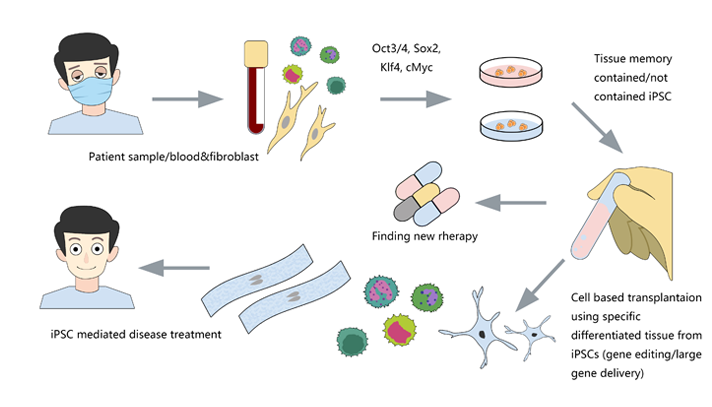
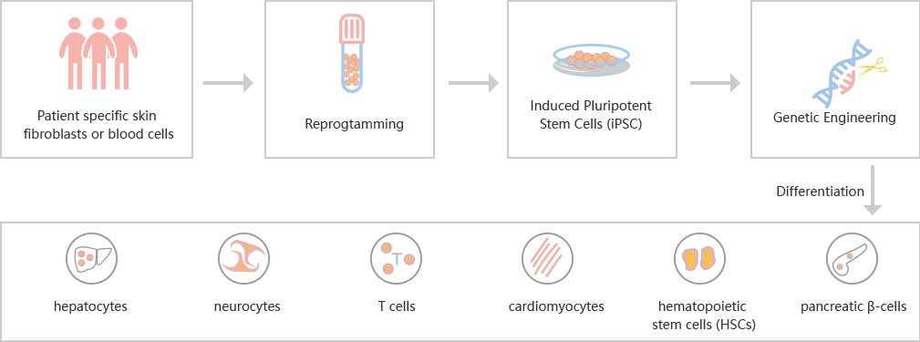
Many serious diseases cannot be cured by medicines, such as heart failure, Late Stage Diabetes, hemophilia, myeloma, End-Stage Cirrhosis, etc. The best method is allogeneic transplantation. However, due to the limited donors and the risk of immune rejection, researchers are dedicated to finding more efficient and safer treatment besides allogeneic transplantation. Induced pluripotent stem cells (iPSCs) can be derived from the body cells of the patients themselves, which eliminates the risk of immune rejection, and has the potential of differentiation into different cells. Transplantation of cells derived from iPSC, such as cardiomyocytes, hepatocytes, neurocytes, T cells, hematopoietic stem cells (HSCs) and islet cells, is possible to solve many medical problems. By CRISPR/Cas9 technology, the mutations that simulating diseases could be introduced into iPSC. Using CRISPR/Cas9 to repair the mutations in iPSC disease models is also a popular application.

CRISPR/Cas9 gene editing in iPSC opens a new window for diseases research

The success rate of gene editing in human iPSC is lower because, unlike tumor cell lines, iPSC does not have the characteristics of chromosomal abnormality and strong ability of DNA repair. CRISPR/Cas9 has the advantages of high efficiency, easy to construct and low toxicity in human cells, so it is the most common method in iPSC genome editing.

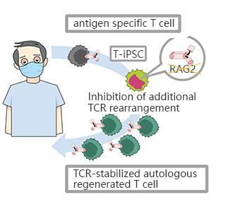
CRISPR/Cas9 mediated RAG2 gene knockout in iPSC, generates CD8αβ - T cells with stable antigen specificity

The limited T cells and the difficulty of proliferation is the main obstacle of T-cell immunotherapy, which can be overcome by using pluripotent stem cells with proliferation and differentiation ability to generate T-iPSC with antigen specificity. Strict antigen specificity is essential for safe and effective T-cell immunotherapy. However, in the process of double-positive CD4/CD8differentiation, the rearrangement of the T-cell receptor (TCR) α chain will lose antigen specificity. This TCR rearrangement was prevented by removing the recombinant enzyme gene (RAG2) in T-iPSCs with CRISPR/Cas9.  Xenotransplantation of CD8αβ-T cells with stable TCR can effectively inhibit tumor growth in disease models. This contributes to a safe and effective T-cell immunotherapy.(Minagawa, Atsutaka, et al.)

CRISPR-UTMcan efficiently transfer gRNA and Cas9 into iPSC by nucleofection. After drug screening, single clones would be generated. Positive clones would be validated by sequencing.


CRISPR/Cas9 was used to repair the point mutation of iPSC disease model derived from an AD patient' cells

Alzheimer's disease (AD) is a progressive and irreversible neurodegenerative disease, which can lead to degeneration of nerve cells and atrophy of brain. It is considered as the most common form of dementia. The A79V mutation of PSEN1 gene can cause Alzheimer's disease. By studying the effect of this mutation on cell phenotype, researchers can further study the pathology of this disease and develop a more effective treatment. The researchers reprogrammed the somatic cells of a patient into pluripotent stem cells (iPSCs), and then replaced the mutated gene with a wild-type sequence. By studying the disease model and the modified iPSC, the effect of the mutation on cell phenotype can be determined, so as to further study the pathological effect of the mutation.(Pires, C., et al.)

With CRISPR-UTM, iPSCwould be co-transfected with gRNA, Cas9 and donor oligo by electroporation. After the DNA DSB caused by the complex of gRNA and Cas9, iPSCs use donor oligo carrying wild-type sequence as a template for homologous recombination repair (HDR) and replace the target sequence with point mutation.


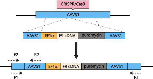
Hemophilia B can be treated by iPSC differentiated hepatocytes with AAVS1 safe harborknockin Coagulation factor IX (F9)

The most common method to treat hemophilia is substitution therapy, but this method has the risk of virus infection, and it is a method that needs lifelong continuous treatment. Gene therapy seems like the only way can cure hemophilia. CRISPR/Cas9 technology can be used for gene therapy of hemophilia. The mutations of coagulation factors, F8 and F9, are the main causes of hemophilia. Previous studies have shown that F9 is a more effective gene therapy target. AAVS1-Cas9-sgRNAplasmid and AAVS1-EF1α-F9 cDNA puromycin donor plasmid were constructed and transferred into iPSC. Human factor IX (hFIX) antigen activity was detected in the culture supernatant. Finally, liver cells differentiated from iPSC were transplanted into NOD/SCIDmice by spleen injection, to cure hemophilia B.(Lyu, Cuicui, et al.)

With CRISPR-UTM, iPSCwould be co-transfected with gRNA, Cas9 and donor vector by electroporation. After drug screening, single clones would be generated. Positive clones would be validated by sequencing.


iPSC induced differentiation, making "autotransplantation" possible

The study of human embryonic stem cells (hESCs) derived from early embryos has been controversial in ethics, and the rejection of differentiated cells derived from hESCs intransplantation has limited its clinical application. Hepatocytes, nerve cells, T cells, cardiomyocytes, hematopoietic stem cells and pancreatic cells can be differentiated from patients' somatic cells (such as fibroblasts) or existing iPSCs.

iPSC Differentiation Process

iPSC reprogramming: Ubigenehas optimized the reprogramming method ——>iPSC induced differentiation: continuous addition of inducing factor in iPSC medium——>Cell validation: genotype, phenotype and cell characteristic analysis

Ubigene's iPSC platform

Ubigene focuses on the optimization of iPSC reprogramming, gene editing and differentiation, and has established a set of mature experimental procedures. With CRIPSR-UTM technology, gene editing in iPSC is much accessible.

If you want to know more, pls click herehttps://www.ubigene.us/about/news/2773.html

[Research highlight] Enhancing p53 pathway can efficiently suppress colon cancer

  Colorectal cancer is the third most diagnosed cancer and leads to the second mortality among cancers worldwide. The first-line chemotherap...