Monday, June 28, 2021

Combination of HCT116 cells and CRISPR/Cas9: A perfect group for colorectal cancer treatment research | Ubigene


 1. Cell information and applications

1.1 Cell information


HCT 116 was isolated from a 48 year old male patient with colorectal cancer in 1979 by M. brattain et al. The cells can form clones in semisolid agarose medium and are tumorigenic in athymic nude mice, forming epithelial-like tumors. So HCT 116 cell line is one of epithelial-like adherent tumor cell lines.


1.2 Cell applications and prospects


(1) To explore the mechanisms by which drugs affect colorectal cancer cell proliferation, migration and invasion. For example: PPARα plays an important role in the migratory activity and the expression of cyp2s1 and CYP1B1 of leukocyte treated HCT116 cells [2];

(2) To explore mechanisms of action in vitro by which drugs affect colorectal cancer cell growth, such as apoptosis and cell cycle alterations. For example: Raddeanin A regulates apoptosis and cycle arrest in human HCT116 cells through the PI3K/Akt pathway [3];

(3)To explore the sensitivity and resistance of drugs to colorectal cancer treatment. For example: Doxorubicin upregulates PD-L1 by inhibiting mir-140 expression in HCT116 cells, thereby rendering tumor cells being resistant to the drug [4];

(4)Development of new cancer-related signaling pathways to provide guidance for clinical treatment of colorectal cancer. For example, to investigate the significance of Notch and Wnt signaling pathways to drug resistance in colorectal cancer cell line HCT116, etc. [5];

(5)Developing new approaches of LncRNA and miRNAs in colorectal cancer treatment. For example: in HCT116 cells, long non-coding RNA of YWHAE competes with miR-323a-3p and miR-532-5p by activating K-Ras/Erk1/2 and PI3K/Akt signaling pathways. It provides a new target site for colorectal cancer treatment. [6];

 2.The application of CRISPR/Cas9 technology in HCT116

As mentioned above, HCT116 cells have been of great research value in the studies on various mechanisms of colon cancer, and the most basic research ideas generally start at the molecular level, that is, gene or protein. Thus, CRISPR/Cas9 technology has emerged in many research topics, and it is nonetheless an irreplaceable advantage in studying the functions of single carcinogenesis gene.


Some researches found that Trpm4 is highly expressed in human colorectal cancer and it seems to be associated with colorectal cancer cell proliferation, cell cycle and invasion. It was found that both tumor cell migration and invasion were indeed reduced by knocking out Trpm4 using CRISPR/Cas9 technology in HCT116 cells, also accompanied by cell cycle alterations [7];


As well as Cell reports that using CRISPR/Cas9 technology, replacing wild-type KRAS with mutant forms rendered the heterozygous mutant HCT116 cells more sensitive to drug treatment [8];


Moreover, by using CRISPR/Cas9 to knockout Tks4 in HCT116 cells, the cells exhibited significant epithelial mesenchymal transition, increased cell motility, and decreased cell-cell adhesion. Therefore found that the Tks4 gene plays an important role in EMT regulation and tumor development [9];


Researchers also found that the CTC1L1142H mutation resulted in impaired telomerase maintenance, by using CRISPR/Cas9 technology to mutate CTC1 in HCT116 cells. It confirmed that the CTC1:STN1 interaction was required for the inhibition of telomerase activity [10].


Therefore, there is no doubt among researchers for the favor of CRISPR/Cas9 technology. Ubigene, by providing the CRISPR/Cas9 technology services, has solved the experimental difficulties for many researchers, such as incomplete cell knockout, unstable cell knock-in, etc.. With our services, more and more researchers can achieve their scientific research objectives smoothly.


 3. Case Studies

3.1 Poing mutation[11]


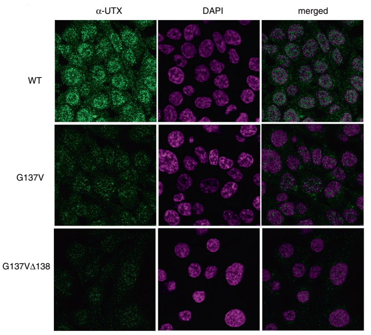
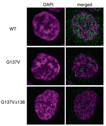
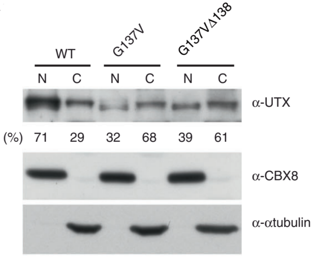
Hiroyuki Kato et al. using CRISPR/Cas9 technology introduced the mutations on UTX gene in HCT16 cells, which is G137V and G137VΔ138. They found that the wild-type UTX, which was originally expressed in the nucleus, greatly decreased in the nucleus, whereas increased in the cytoplasm in both mutant cells. As shown in Figure A to C: A, B were the immunofluorescence figures, C was the Western blot results. The results of co-immunoprecipitation are shown in fig.d, it is a novel regulatory mechanism of UTX. Also, the article also further revealed the importance of UTX interaction with MLL3/4 complex in cancer formation (ASH2L, PTIP and PA1 are components of MLL3/4 complex) .
3.2 Knockout [12]


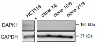
CRISPR/Cas9 gene-editing technology is used to obtain HCT116 cell line with DAPK1 deletion by Sara Steinmann et al, and finally revealed the effect of DAPK1 on the invasion of colorectal cancer.


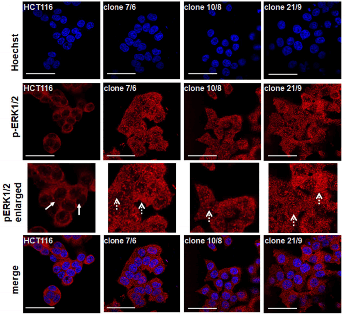
After Western Blot verification, three DAPK1 gene knockout monoclonal cells (Fig. A) were obtained by Sara Steinmann et al. Immunofluorescence assays detected pERK1/2 mainly in the wild-type HCT116 cytoplasm. However, in all three knockout cells, pERK1/2 was significantly expressed in the nucleus. (Fig. B)


Because the chorionic allantoic membrane model (CAM) experiment is a classic in vivo model of angiogenesis, the researchers transplanted DAPK1 knockout clone and wild- type HCT116 cells to chicken CAM, and cultured in eggs for 5 days. It was found that the absence of DAPK1 led to the change of growth pattern and enhancement of tumor bud in CAM ( Fig. C)


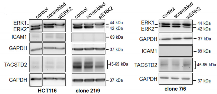
In addition, the team used a rat brain 3D model in vitro to find that tumor cells had more proliferation in chicken embryonic organs, and the invasion ability was also enhanced. DAPK deficient HCT116 cells showed more diffuse tumor cells and preferentially accumulated in the liver, heart, and brain of the chicken embryo (Fig. D). Finally, the researchers found that DAPK1-ERK1 signaling pathway is involved in the metastasis of CRC (Fig. E).


3.3 Knock-in [13]


BAX is one of the members of pro-apoptotic Bcl-2 gene family, which plays an important role in mitochondria dependent apoptosis initiation. R Peng et al have knocked in five mutation sites in BAX-KO HCT116 cells, which are located in the BAX gene and interact with other members of Bcl-2.


It has been reported that BAX-KO HCT116 cells do not undergo apoptosis by the stimulation of sulindac, a steroidal drug. R Peng et al found that knock in WT BAX in BAX-KO HCT116 cells can restore sulindac, and can restore HCT116 apoptosis caused by TRAIL. After knocking in the K21E and D33A mutations, BAX mediated apoptosis was completely restored; Knocking-in D68R and S184V mutations only partially recovered, while knocking in L70A / D71A mutation caused less apoptosis.
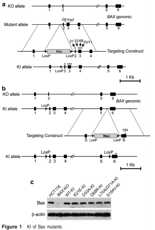
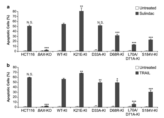

In this study, the target gene in the target cell is knocked out, and then knock in the target gene containing the mutation site and WT target gene (contrast), then we can clearly understand the specific site relationship between the protein expressed by the target gene and other proteins interacting with it, It is a good method to verify whether the predicted sites obtained by bioinformatics analysis really play a role in protein-protein interactionUbigene also provides the corresponding service convenience for scientific researchers. While enjoying the gene KI cell line service, free knockout cells can be obtained which meets the various research needs.

Ubigene CRISPR-UTM technology with 5000 successful experiences makes our gene-editing services more trustworthy! We have promotions for gene-editing services now: KO cell line services as low as 3780 USD KI/PM cell line services as low as 8480 USDgRNA plasmids as low as 80 USD

Reference:

[1]Bray, Freddie et al. “Global cancer statistics 2018: GLOBOCAN estimates of incidence and mortality worldwide for 36 cancers in 185 countries.” CA: a cancer journal for cliniciansvol. 68,6 (2018): 394-424. doi:10.3322/caac.21492

[2]Khor CY, Khoo BY. PPARα plays an important role in the migration activity, and the expression of CYP2S1 and CYP1B1 in chrysin-treated HCT116 cells. Biotechnol Lett. 2020;42(8):1581-1595. doi:10.1007/s10529-020-02904-

[3]Meng C, Teng Y, Jiang X. Raddeanin A Induces Apoptosis and Cycle Arrest in Human HCT116 Cells through PI3K/AKT Pathway Regulation In Vitro and In Vivo. Evid Based Complement Alternat Med. 2019;2019:7457105. Published 2019 May 26. doi:10.1155/2019/7457105

[4]Naba NM, Tolay N, Erman B, Sayi Yazgan A. Doxorubicin inhibits miR-140 expression and upregulates PD-L1 expression in HCT116 cells, opposite to its effects on MDA-MB-231 cells. Turk J Biol. 2020;44(1):15-23. Published 2020 Feb 17. doi:10.3906/biy-1909-12

[5]Kukcinaviciute, Egle et al. “Significance of Notch and Wnt signaling for chemoresistance of colorectal cancer cells HCT116.” Journal of cellular biochemistry vol. 119,7 (2018): 5913-5920. doi:10.1002/jcb.26783

[6]Bjeije, Hassan et al. “YWHAE long non-coding RNA competes with miR-323a-3p and miR-532-5p through activating K-Ras/Erk1/2 and PI3K/Akt signaling pathways in HCT116 cells.” Human molecular genetics vol. 28,19 (2019): 3219-3231. doi:10.1093/hmg/ddz146

[7]Kappel, Sven et al. “TRPM4 is highly expressed in human colorectal tumor buds and contributes to proliferation, cell cycle, and invasion of colorectal cancer cells.” Molecular oncology vol. 13,11 (2019): 2393-2405. doi:10.1002/1878-0261.12566

[8]Szeder, Bálint et al. “Absence of the Tks4 Scaffold Protein Induces Epithelial-Mesenchymal Transition-Like Changes in Human Colon Cancer Cells.” Cells vol. 8,11 1343. 29 Oct. 2019, doi:10.3390/cells8111343

[9]Burgess, Michael R et al. “KRAS Allelic Imbalance Enhances Fitness and Modulates MAP Kinase Dependence in Cancer.” Cell vol. 168,5 (2017): 817-829.e15. doi:10.1016/j.cell.2017.01.020

[10]Gu, Peili et al. “CTC1-STN1 coordinates G- and C-strand synthesis to regulate telomere length.” Aging cell vol. 17,4 (2018): e12783. doi:10.1111/acel.12783

[11]Kato, Hiroyuki et al. “Cancer-derived UTX TPR mutations G137V and D336G impair interaction with MLL3/4 complexes and affect UTX subcellular localization.” Oncogene vol. 39,16 (2020): 3322-3335. doi:10.1038/s41388-020-1218-3

[12]Steinmann, Sara et al. “DAPK1 loss triggers tumor invasion in colorectal tumor cells.” Cell death & disease vol. 10,12 895. 26 Nov. 2019, doi:10.1038/s41419-019-2122-z

[13]Peng, R et al. “Targeting Bax interaction sites reveals that only homo-oligomerization sites are essential for its activation.” Cell death and differentiation vol. 20,5 (2013): 744-54. doi:10.1038/cdd.2013.4

Wednesday, June 16, 2021

Will CRISPR/Cas9 technology lead to new development of cell genetic modification? Ubigene


Since the world’s first CRISPR/Cas9 clinical trial was carried out in West China Hospital in 2016, gene therapy for cancer has made many gratifying breakthroughs with the help of CRISPR/Cas9, and the breakthrough of CRISPR/Cas9 also attracted everyone's attention. Ubigene has been committed to CRISPR/Cas9 system in cell gene-editing experiments. We are ahead of other companies and promise to guarantee 100% no protein residue in 5000 gene knockout experiments. We also pay attention to the progress of CRISPR/Cas9 system in the field of cancer. Here are a few cases of cancer research using CRISPR/Cas9 system (the following cells are all successful cases of Ubigene):

Gene editing of Huh-7 cell line——Boost coronavirus, drug metabolism and cancer research

Huh-7 cell line was established in 1982 by Nakabayshi, H. and Sato, J. it is epithelioid and highly heterogeneous. In order to overcome the shortcomings of primary hepatocytes in drug metabolism research, a CRISPR/Cas9 gene modified human hepatocyte line was developed,which studies the effects of gene variation on drug metabolism. Cancer stem cells (CSCs) are closely related to the occurrence and metastasis of cancer. They have the ability of self-renewal and unlimited proliferation, and are the key factors in the development of cancer. Researchers found that the expression of androgen receptor was very high in liver cancer tissues, and was related to Nanog. CRISPR/Cas9 technology was used to knock GFP into Huh-7 cells Nanog, revealing the gender difference in incidence rate of hepatocellular carcinoma, and providing a possible way for the suppression of axons in liver cancer treatment. 

click here for the full article

Gene editing CT26.WT cell line——A magic way for colon cancer research and treatment

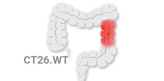
CT26.WT cell is an invasive mouse colon cancer cell line. By using CRISPR/Cas9 system to edit gene in this cell line, we can generate single or multiple gene knockout, mutation correction or reporter gene insertion transgenic cells. It has a wide range of applications in the study of cancer markers, revealing the mechanism of drug resistance, cancer treatment, cell death and other fields. The researchers used CRISPR/Cas9 system to knock out ATG7 in mouse melanoma cell line B16F10, mouse colon cancer cell line MC38 and mouse colon adenocarcinoma cell line CT26 to study the role of autophagy in the proliferation of mouse cancer cells, and finally revealed the effect of autophagy destruction on immune reactive tumors.

click here for the full article


H1299 cells——A tool of studying CRISPR gene therapy for cancer mutation

As an immortalized cell line, H1299 can divide infinitely. The unique feature of this cell line is the lack of P53 protein expression. As a disease model, H1299 cell line plays a very important role in understanding the basic biology of disease, understanding how mutations affect drug response or drug resistance, understanding drug reactivity, target recognition, verifying the mechanism of differences, and even stratification of patients. In one study, researchers used CRISPR/Cas9 technology to construct BCAR1 gene knockout on lung adenocarcinoma cell lines (ncl-h1975 and ncl-h1299), and showing that BCAR1 promoted the proliferation and cell growth of lung adenocarcinoma by up regulating POLR2A.

click here for the full article

Ubigene has been working to allow CRISPR/Cas9 technology to be better developed and applied, and our independently developed CRISPR-U™ Technology is 10-20 times more efficient than traditional gene editing, also the breakthrough patented technology for gene modification in vitro and in vivo. Moreover, Ubigene uses CRISPR-U™ to successfully edit genes from more than 100 cell lines. Our leading technology with abundant experience makes our gene editing services more trustworthy!

Wednesday, June 9, 2021

Point mutation cell line generation--Ubigene Weekly Q&A


Nowadays, many researches indicate that CRISPR/Cas9 is becoming the optimal tool to construct the models of human diseases, such as human pluripotent stem cells and tumor cell lines. With modern sequencing, CRISPR/Cas9 can introduce precise point mutations (homozygous or heterozygous) in various cell lines and enables to construct human disease models. This is highly advantageous for studying both the therapy and the underlying genome of the diseases.


We also received a few questions this week about introduction of point mutation in cells using CRISPR/Cas9. Below are the Q&As for this week.


Q1 How can I use CRISPR/Cas9 technology to study gene point mutation cell lines?

A

The specific mutations can be achieved by CRISPR/Cas9 efficient introduction of targeted double-strand breaks (DSBs), repaired using DNA repair donor (e.g., single-stranded oligo DNA nucleotide, ssODN) under homology-directed repair (HDR) system. Importantly, with the KI cassette based on the CRISPR/Cas9 system, we can also selectively introduce mono-allelic and dual-allelic sequence changes, which are heterozygous and homozygous point mutations to construct pathogenic disease models.


Homozygous point mutations require gRNAs to target positions close to the target mutation site, whereas heterozygous point mutations can be achieved by distance-dependent incorporation of suboptimal mutations or by using mixed repair templates.


So, the introduction of point mutations in cell lines mediated by CRISPR/Cas9 technology is very useful for the studies on the gain of function or loss of function.


Q2 Hi there, I want to construct a gene point mutated Hep-G2 cell line. For reference, could you tell me the estimated timeline for the whole experiment?

A

For endogenous point mutation, it’s a bit challenging to introduce in Hep-G2 because the proliferation rate for Hep-G2 is relatively slow and the single-cell colony formation rate is relatively lower than other common cell line models. If everything goes smoothly, the whole process takes about 3-4 months. Because it is difficult and time consuming, you could consider outsourcing the experiment.


Ubigene is experienced in successfully modifying Hep-G2 cell line and if you order point mutation cell line generation services from Ubigene, we will give you the KO cell line for free, making your experiment and researches more convenient.


Q3 Hi there, I want to introduce a point mutation (change of an amino acid) in the target gene using CRISPR/Cas9 technology, and I want to do cell transfection and single-cell clone isolation in my own lab. I have searched many articles and checked some information from addgene, found that I need to construct a donor DNA to achieve the point mutation in the cells. How to design the constructs and how much budget is generally required?

A

The donor DNA is not as complicated as you think to construct. A donor comprises a sequence carrying the mutation and upstream and downstream sequences (each about 1-2Kb long) for the purpose of homologous recombination. Or you could also use oligo which has shorter upstream and downstream sequences (about 100-200bp) to achieve the point mutation.

For the question of budget, the regular price for a set of point mutation constructs and oligo is about 770 USD in Ubigene.

Above are the Q&As of the week.

Feel free to contact us if you have any similar questions.


Ubigene makes genome editing easier!

Tuesday, June 1, 2021

CRISPR/Cas9 Stable Cell Line / Virus Packaging service $500 cash back on every 5K USD spent, Order now to get a $800 coupon, which can be used for a variety of cell assay services | Ubigene Biosciences


Ubigene Red Cotton™ · gRNA plasmid bank, Over 10,000 in-stock gRNA plasmids as low as $80


Thursday, May 20, 2021

Ubigene Biosciences Gene Point Mutation/ Knockin Cell Line service , Comes with 2000 USD Cell Assay Coupon


Gene Knockout cell line, 100% WB guarantee Only 3780 USD | Ubigene Biosciences


Friday, May 14, 2021

Application of KO cells | Ubigene Biosciences


CRISPR (Clustered Regularly Interspaced Short Palindromic Repeats), the term refers to a series of repetitive patterns in the DNA of bacteria and archaea that were extensively studied by Spanish scientist Francis Mojica in the ‘90s. These patterns are the basis of a primitive immune system that bacteria use to ‘remember’ the DNA of viral invaders by incorporating the DNA sequence of the virus within the CRISPR patterns. The Cas9 protein is then able to recognize the DNA sequence stored within CRISPR patterns and cut any DNA molecules with a matching sequence. Since 2012, the CRISPR/Cas systems was developed into easy-to-use and robust genome editing tool for basic research and clinics.


Three common strategies have been developed for genome editing with the CRISPR/Cas9 platform:


(1)The plasmid‐based CRISPR/Cas9 strategy, where a plasmid is used to encode Cas9 protein and sgRNA, assembles Cas9 gene as well as sgRNA into the same plasmid in vitro. However, the encoded plasmid needs to be introduced inside the nucleus of target cells, which is a key challenge in this system.

(2)Direct intracellular delivery of Cas9 messenger RNA (mRNA) and sgRNA, the greatest drawback of which lies in the poor stability of mRNA, which results in transient expression of mRNA and a short duration of gene modification;

(3)Directly delivery of Cas9 protein and sgRNA, which has several advantages, including rapid action, great stability, and limited antigenicity. Among the versatile CRISPR-based tools, CRISPR/Cas9 is the most widerly used one for studying gene functions. It has boost the generation of knockout cell lines for different applications.


  Knockout cellular model applications:

There is a significant challenge in translating the abundance of genetic information. To date, it is possible to identify the role of genes, to understand basic biology, as well as linking to the role of mutations for understanding disease pathogenesis. A Knockout (KO) cell lines are excellent model systems to do this. The advantage of cell lines is the ability to use gene-editing to construct isogenic cell line pairs, where a mutant model can be probed alongside a wild-type control. The following are potential examples of how to get the most out of your research using KO cell lines:


1. Antibody specificity validation

2. Identify pathway players

3. Identify drug targets

4. Build disease models

5. Develop therapeutic tools


1. Antibody specificity validation


The research community believes that the use of excellent research tools impacts significantly the quality of research delivered. Antibodies are one of the most commonly used reagents in life-sciences, but despite their widespread use, there are no standard guidelines for how these invaluable biological tools should be validated prior to use. For example, poorly characterized antibodies may yield non-specific results, which are difficult to replicate even when the only difference is the antibody's production lot. However, the bar for antibody validation is rising and there are calls for the industry standard to show application-specific validation using KO cell lines. So, KO cell line has wide application in antibody validation. The parallel use of wildtype and KO cell lines provides a valuable tool to control for research reagents quality.


2. Identify pathway players


Often there are multiple related, but distinct molecules and processes present in a pathway. Gene-edited cell lines are a well-established tool to probe the role of a particular gene or mutation. For example, JAK1 is essential for signaling for certain cytokines and is thought to play a role in tumor metastasis. It is also known to have a functional and physical association with INF gamma receptors. When ligands bind with receptors, the receptor-associated JAKs become activated, leading to the recruitment of specific STATs (signal transducer and activator of transcription) from the cytoplasm. These STATs then become JAK substrates. Activated STATs are released from the receptor, which then translocates to the nucleus to bind to specific enhancer elements. These then affect the expression of target genes. The role of JAK1 in STAT phosphorylation can be confirmed by creating a frameshift mutation in the JAK1 gene and the KO cell line do not allow phosphorylation of STAT and this could be verified with westernblot analysis.


3. Identify drug targets


Pharmacological interventions are commonly used to interrogate pathways. For example, the chemotherapeutic 6-thioguanine (6-TG) is used to probe the effect of DNA damage response. To maintain genomic integrity, cells are equipped with a myriad of mechanisms that are each specific for different types of damage. The efficiency of these DDR pathways plays an essential role in the effectiveness of cytotoxic treatments. 6-TG is a growth inhibitory antimetabolite that requires an active DNA mismatch repair (MMR) system to be effective. One study showed that HPRT+ cells are sensitive to 6-thioguanine (6-TG), which can be converted to the nucleotide form by HPRT and incorporated into DNA by DNA polymerase, killing cells by a process involving postreplicative mismatch repair. The strategy is to transfect cells with two plasmids that express respectively a HPRT guide RNA and a guide RNA for the gene of interest. Cas9 can be expressed from the gene on a separate plasmid, a plasmid carrying the HPRT gRNA or integrated into the chromosome (if such a cell line is already available). If a cell becomes resistant to 6-TG, it would suggest that this cell should also be competent to target the gene of interest as long as the gRNA is effective. Thus if the targeted gene is not altered in the resulting 6-TG resistant cells, it would suggest that the guide RNA is ineffective. On the other hand, if no 6-TG resistant cells can be obtained by co-targeting, it would suggest that the gene of interest might be essential. Thus KO cell lines that are devoid of key proteins are used to confirm the target of the 6-TG.


4. Build disease models


After identifying mutations of interest in a patient population, the next steps are to probe the biological mechanisms that result in the disease phenotype or the response to treatment. KO cell lines, combined with simple molecular biology techniques, allows the researcher to rescue the gene of interest to restore phenotype and complement the Knockout cell line with a functional mutation, such as a point mutation observed in a patient population. One example of disease modeling is MAGEC2 knockout in melanoma cancer cell line. MAGEC2, a member of the type I melanoma-associated antigen family, is expressed in a wide variety of cancer types but not in normal somatic cells.  Reserchers generated MAGEC2-knockout A375 melanoma cell lines using the CRISPR/Cas9 system. They found that knockout or knockdown of the MAGEC2 gene sensitized melanoma cells to tumor necrosis factor-α-induced apoptosis. Their study sheds a light on the molecular pathway by which MAGEC2 promotes tumor development.


Earlier studies reported that Nuclear Factor Erythroid 2-Related Factor (NRF2) is a master regulator of 100–200 target genes involved in cellular responses to oxidative and/or electrophilic stress. NRF2 is also known to regulate the expression of genes involved in protein degradation and detoxification, and it is negatively regulated by Kelch-like ECH-associated protein 1 (KEAP1), a substrate adaptor for the Cul3-dependent E3 ubiquitin ligase complex. One research group used CRISPR/Cas9 to disable the NRF2 gene in lung cancer cells by disrupting the NRF2 nuclear export signal (NES) domain; phenotypically, the protein is largely blocked from transiting into the nucleus after translation. Cells with this gene knockout were found to have a reduced proliferation phenotype and are more sensitive to chemotherapeutic agents, such as cisplatin and carboplatin. They also found that homozygous knockout cells proliferate at a slower rate than the wild-type cells, even in the absence of drug treatment.


5. Develop therapeutic tools


During the past few years, the team represented by China and the United States have conducted a series of clinical trials of gene editing, such as producing more effective CAR T cells for the treatment of cancer and the knockout of the erythroid-specific enhancer of BCL11A to upregulate gamma globulin in autologous erythroid HSCs as a potential therapy for sickle cell disease and β-thalassemia. To date, several ongoing clinical trials applying the concept of PD-1 knockout autologous T cells to treat cancers, including prostate cancer (NCT02867345), esophageal cancer (NCT03081715), and renal cell cancer (NCT02867332). These trials are considered proof-of-concept studies to apply the in vitro CRISPR/Cas9 gene knockout technique in cancer therapy.


In summary, KO cell lines are a versatile tool for helping us understand basic biology, as well as identifying the role of mutations in disease pathogenesis. Ultimately, this has led to advances in disease treatment and patient prognosis.


Reference:

Enriching CRISPR-Cas9 targeted cells by co-targeting the HPRT gene. Nucleic Acids Res. 2015. 43(20): e134.

Establishment of MAGEC2-knockout cells and functional investigation of MAGEC2 in tumor cells. Cancer Sci 2016. 107(12):1888-1897.

Functional Gene Knockout of NRF2 Increases Chemosensitivity of Human Lung Cancer A549 Cells In Vitro and in a Xenograft Mouse Model. Molecular Therapy: Oncolytics. 2018 11:75-89.

Applications of genome editing technology in the targeted therapy of human diseases: mechanisms, advances and prospects. Signal Transduct Target Ther .2020 Jan 3;5(1):1.

Monday, May 10, 2021

[One-Stop Services] Cell Assay Services | Ubigene


What needed to deal with after the gene-editing experiment for a cell is done?
A subsequent series of cell assays are important as well! But!
 No equipment to do the cell-based assays for phenotypic analysis?
 Too messy and huge workload for data analysis?
 No appropriate service provider found?


No worries! Let Ubigene rescues you!
Ubigene have now launched a series of new services - Cell assays
Providing One-Stop Services from cell line generation to cell phenotypic analysis
Ubigene can help save your time from tedious, repetitive experiments and obtain
the experimental data for you.


Ubigene has been receiving the feedback from customers and trying our best to reach the requirements. So, we released the cell phenotypic analysis services, covering a wide range of popular cell-based assays such as assays for cell proliferationcell apoptosiscell migration and invasioncell cyclecytotoxicitymarker protein expression levels, and cytokine secretion/enzyme activity (ELISA), and so on. Feel free to contact us for more information and ordering.

Here is the big promotion for you!

As long as you have ordered any KO / Overexpression / Knockdown Cell Line project from Ubigene, you can enjoy the promotional price for the following all-in-one package of cell assays.

Promotional package: cell subculturing, cell proliferation assay, cell migration or invasion assay, and cell cycle assay.


Thursday, May 6, 2021

Cell migration and invasion-Principles and Methods | Ubigene

Cell migration and invasion

Cell migration, also known as cell crawling, or cell movement, refers to the movement of a cell after receiving a migratory signal or sensing a gradient of certain substances. Cell migration is an alternating process of pseudopodia extension at the cell head, the establishment of new adhesions, and retraction of the cell body tail in a spatiotemporal manner. Cell migration, one of the basic functions of normal cells, is a physiological process of normal growth and development of the organism, and a ubiquitous form of movement of living cells. Cell migration is implicated in processes such as embryonic development, angiogenesis, wound healing, immune responses, inflammatory responses, atherosclerosis, and cancer metastasis.

Cell invasion refers to the ability of cells to migrate from one area to another through the extracellular matrix. Cell invasion is a response of normal and cancer cells to chemical and mechanical stimuli. Before migrating to new areas, the extracellular matrix is degraded by proteases within the cell. Cell invasion often occurs during wound repair, vascularization and inflammatory response as well as abnormal tissue infiltration, tumor cell metastasis, and so on.


Technical Details
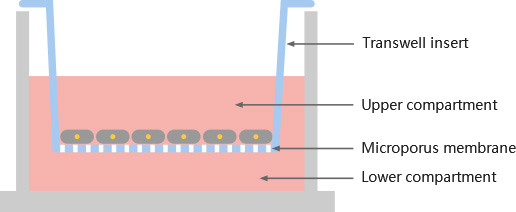
Transwell assay is a commonly used to detect the migration ability and invasion ability of cells. Transwell assay, as its name, that is, the compartment with densely packed small holes is put into a well plate (commonly a 24 well plate) . The cell suspension is added to the compartment, which is placed in a 24 well plate with complete medium added. The cells can pass through the holes in the compartment by deformation and run to the outside of the more nutritious compartment and attach to the outside. By staining and counting the cells on the outside of the compartment, it can tell the strength of the migration and invasion ability of the cells.The basic principle of Transwell is to put the small compartment into a culture plate, inside the small compartment called upper compartment, inside the culture plate called lower compartment. The upper and lower culture fluids are separated by a polycarbonate membrane, upper compartment is added by upper culture fluid, lower compartment is added by lower culture fluid. Add the cells to the upper compartment, and because the membrane is permeable, the components in the lower compartment can affect the cells within the upper compartment, so that the effects of the components of culture medium in the lower compartment on cell growth, motility, and so on can be studied.

Tumor migration assay is the study of the migratory ability of tumor cells or, in specific circumstances. Commonly 8.0, 12.0 µ m membrane is used, adding the tumor cells to the upper compartment, and FBS or some specific chemokines are added in the lower compartment. Tumor cells will migrate towards the lower compartment with high nutrient content. Counting the number of cells entering the lower compartment can indicate the migration ability of tumor cells.

Tumor invasion assay is the study of the invasive ability of tumor cells or, in specific circumstances. Unlike the tumor cell migration assay, a layer of Matrigel is additionally added above the polycarbonate membrane to mimic the extracellular matrix. Tumor cells are added to the upper compartment, and if tumor cells want to enter the lower compartment, they must first secrete matrix metalloproteinases (MMPs) to pass through the polycarbonate membrane by digesting the Matrigel. Counting the number of cells that enter the lower compartment can indicate the invasive ability of tumor cells.

Case study

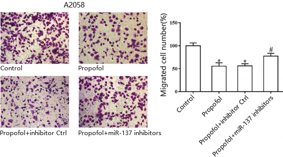
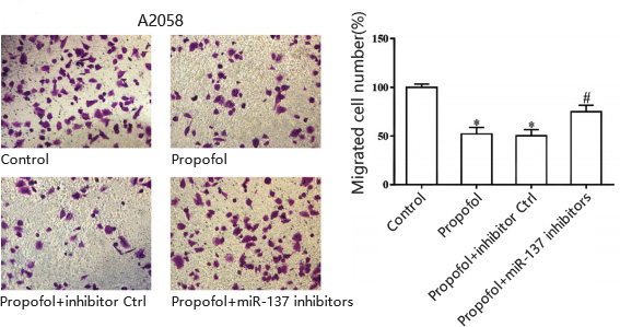
A.Cell migration for human skin melanoma cell line A20578

B.Cell invasion for human skin melanoma cell line A20578


Reference
Yu, Hong et al. “Propofol suppresses proliferation, invasion, and migration of human melanoma cells via regulating microRNA-137 and fibroblast growth factor 9.” Journal of cellular physiology vol. 234,12 (2019): 23279-23288. doi:10.1002/jcp.28896


Service turnaround and deliverables

Service turnaround

5~10 business days, depending on cell growth and experimental design


Customer provides

cell lines, drugs, drug treatments and experimental conditions parameter settings;


Deliverables

cell lines, drugs, drug treatments and experimental conditions parameter settings;


Other methods to test cell migration and cell invasion


Wednesday, May 5, 2021

Cell Proliferation-Principles and Methods | Ubigene

Cell Proliferation

Cell proliferation is one of the important physiological functions of living cells and is an important life characteristic of organisms. Cell proliferation is the basis of organism growth, development, reproduction as well as heredity.


Technical Details


Normal cells are metabolically vigorous and their mitochondria contain succinate dehydrogenase, which can reduce tetrazolium salts (e.g., MTT, XTT, WST-1, and WST-8, etc.) to purple crystalline substances that deposit around the cell. Then we can use a microplate reader to read the OD value (optical density), thus detecting the status of cell proliferation.

Cell counting kit-8 (also known as CCK-8) is a rapid and highly sensitive assay based on WST-8 (Water Soluble Tetrazolium-8) and widely used for cell proliferation analysis.

The mechanism is: WST-8 [chemical name: 2-(2-Methoxy-4-nitrophenyl)-3-(4-nitrophenyl)-5-(2,4-disulfophenyl)-2H-tetrazolium Sodium Salt] is reduced by dehydrogenases in cells under the reaction of the electron carrier 1-methoxy-5-methylphenazinium sulfate dimethyl ester (1-methoxy PMS) to a highly water-soluble yellow formazan product (formazan dye).The number of formazans generated directly correlates to the number of living cells.This property can therefore be exploited to directly carry out cell proliferation assays.The more rapidly the cells proliferate and the more cells proliferate, the darker the color is. For the same cells, there is a linear relationship between the intensity of the color and the number of cells.


Case study


Reference
Deng, Danni et al. “p62 acts as an oncogene and is targeted by miR-124-3p in glioma.” Cancer cell international vol. 19 280. 6 Nov. 2019, doi:10.1186/s12935-019-1004-x


Service turnaround and deliverables

Service turnaround

5~10 business days, depending on cell growth and experimental design


Customer provides

cell lines, and experimental conditions parameter settings


Deliverables

raw data, analytical results, experimental reports

Other methods to detect cell proliferation


[Research highlight] Enhancing p53 pathway can efficiently suppress colon cancer

  Colorectal cancer is the third most diagnosed cancer and leads to the second mortality among cancers worldwide. The first-line chemotherap...