Wednesday, April 28, 2021

CRISPR gene-editing | Best Choice for Research | Ubige


shRNA vs gRNA:


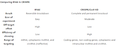
RNAi technology used to be popular in biological research, especially in high-throughput genetic screening, gene function study, clinical treatments for antivirus and cancers. However, the RNAi technology has been questioned because of its off-target effect. In recent years, the emerging CRISPR gene editing technology is widely used in almost every biological field. Because of its high specificity, low off-target effect and wide application prospects, CRISPR is replacing RNAi as the best choice for most research.


What RNAi might cost you?

One of the biggest limitations of the RNAi silencing method is that it suffers from High off-target effects. RNAi might silence unintended RNA targets resulting in modified phenotypes and therefore influence gene function screening experiments. There are three reasons for this high off-target effect:

· siRNA may compete with the intrinsic microRNA in RISC complex, resulting in affecting the function of microRNA and the expression regulation of other genes;
· siRNA also targeted sequences with limited complementarity;
· siRNA may interact with the 3'UTR of unintended RNA, leading to its degradation.

Other than high-off target effect, issues that researchers are facing with are as followings:
· shRNA fails to show any reduction in protein expression level;
· No suppression but overexpression of target genes;
· Transcripts with high turnover are difficult to silence;
· Non-coding sequences are difficult to silence.

CRISPR has replaced RNAi as the best choice for most research applications:

Researchers conducted parallel experiments with shRNA libraries and CRISPR gRNA libraries, which proved that the efficiency and reliability of using gRNA libraries to screen genes were much higher than that of shRNA libraries.

In addition to high efficiency and high true positive rate, the off-target effect of CRISPR is very low. Because its specificity depends on two aspects, one is the base pairing between gRNA and target DNA, the other is that the Cas9-gRNA complex only binds to the PAM sequence in the genome. Only when these two conditions are met at the same time can Cas9 cause DSB.

Although RNAi was adopted as a gene silencing technique first, CRISPR has surpassed RNAi in popularity due to several advantages made possible by advancements that have refined CRISPR technology. The main reason for the popularity of CRISPR is it's specific yet versatile nature. At the same time, the choice remains with the user to perform knockouts, knock-ins, or knockdown experiments, making CRISPR extremely versatile.

Combine RNAi with CRISPR technology:
In some cases, CRISPR and RNAi can work with each other. For instance, it is very rare for any drug to achieve 100% inhibition of the target gene, so some scientists believe that a more rigorous and ideal experiment design is to use shRNA to validate a phenotype observed by CRISPR technology.


Ubigene Signature Services:
Ubigene has an advanced platform for molecular and cellular experiments. Our experienced technical team can design high-score shRNA and high-efficiency gRNA for you to meet the needs of your RNAi and CRISPR knockout experiments.

· gRNA and shRNA clones of different species;
· Virus packaging for gRNA or shRNA, including lentivirusesadenoviruses and AAVs;
· Custom stable cell lines with gene knockout or knockdown;
· Gene knockout and knockdown zebrafish, and related downstream analytical services.
References

· Combined shRNA over CRISPR/cas9 as a methodology to detect of-target efects and a potential compensatory mechanism
· CRISPR knockout screening outperforms shRNA and CRISPRi in identifying essential genes
· Systematic comparison of CRISPR/Cas9 and RNAi screens for essential genes
· Choosing the Right Tool for the Job: RNAi, TALEN, or CRISPR

Sunday, April 25, 2021

Knockout cell line | circRNA' function in Gene Regulation | Ubigene

 CRISPR/Cas9 Mediated circRNA Knockout, Reveals Its Roles in Gene Regulation


circRNAs are circular noncoding RNAs formed by reverse splicing of pre-mRNAs. circRNAs were firstly found in viruses in the 1970s. However, due to the extensive use of the method for enrichment of poly (A) (no 5 'and 3' ends of circRNA) in the early RNA library preparation, and the calculation algorithm that RNA-seq reading requires linear alignment with genome, a large number of circRNA information was omitted, which led to the belief that circRNA is just a byproduct of miss splicing. 

With the development of high-throughput sequencing technology and bioinformatics, thousands of circRNA have been found, and more and more basic researches related to circRNA have been done. A large number of studies have shown that circRNA is endogenetic, abundant, conservative and stable in mammalian cells, and often shows tissue or space-time specificity. It can participate in the regulation of cell growth and development, as well as the occurrence and development of diseases through a variety of mechanisms. Therefore, in recent years, circRNA has become popular in the field of non-coding RNA research.

Relationships between circRNAs and diseases

At present, the most studied is the relationship between circRNAs and tumors. Some circRNAs promote tumor formation, such as circPvt1 in squamous cell carcinomas of the head and neck, cirs-7 (CDr1as) in colorectal cancer, esophageal squamous cell carcinoma and hepatocellular carcinoma. Some circRNAs suppress tumors, such as circsMARCA5 and circ-SHPRH in glioblastoma. Some circRNAs may play different roles in different tissues or cells, such as circHiPK3, which is a proto-oncogene in rectal cancer, but suppresses cancer cells in bladder cancer.

In addition to cancers, circRNA has been found to be closely related to diabetes, cardiovascular disease, chronic inflammation and nervous system diseases. It is believed that with the development of biotechnology and more in-depth researches on circRNA, the formations and mechanisms of circRNAs can be identified. circRNAs can play important roles in disease prevention, diagnosis and treatment discovery.

The engineering and regulation of circRNA help disease studies and treatment discoveries

circRNA involves many complex functions, so how to study its functions? Similar to protein-coding genes, the most common methods are knockout, knockdown (RNA interference) or overexpression of the circRNA.

CRISPR/Cas9 mediated circRNA knockout, reveals the mechanism of its regulation on tumor formation

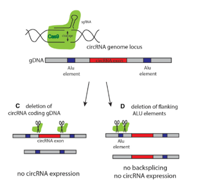
circRNA knockout refers to editing at the level of DNA to achieve the purpose of a complete knockout. The most commonly used method is to design two gRNAs at both ends of the circRNA exon to knockout the whole cyclized exon sequence. Although this strategy can knockout the circRNA, it will also affect the parent gene encoding the protein, and the study on its function is not ideal.

More and more researchers have given up using this strategy to knockout the circRNA because of its great influence on the parent gene. The ideal method is to knockout the loop forming elements (Alu) in the flanking intron of the exon, so as to destroy the circRNA loop forming without affecting the expression of the coding gene. However, it is difficult for many researchers to master the logic of designing the targeting strategy.

The most common strategy for circRNA knockout


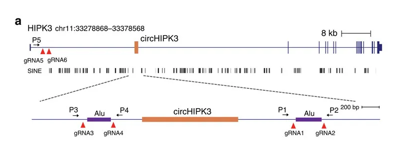
Taking circ-HIPK3 as an example, circ-HIPK3 is a kind of circRNA rich in human cells, which can combine with a variety of miRNAs as a regulator of cell growth and affect the formation of tumors. In order to verify how circ-HIPK3 forms into circle, it is necessary to find the loop forming elements in flanking intron. A pair of sgRNA is designed for the two Alu elements predicted at upstream and downstream respectively. The predicted loop forming elements are knockout by CRISPR/Cas9 system to detect whether the expression of circRNA changes. After PCR and RT-qPCR verification, it was found that the expression of circ-HIPK3 was significantly down-regulated after knockout of downstream loop forming elements, while the expression of circ-HIPK3 was not decreased but increased after knockout of upstream loop forming elements. It was speculated that there were too many loop forming elements in the upstream and the prediction was not accurate. In order to further verify the RNA circulation driven by other elements, the large fragment of intron in the upstream of the element was knockout by co-injection of gRNA3 or gRNA4 with gRNA5 or gRNA6. RT-qPCR results showed that the expression of circ-HIPK3 decreased, indicating that other loop forming elements of circ-HIPK3 exist.

Loop-forming elements predicted by the CRISPR/Cas9 system


Ubigene is experienced in designing strategy of knockout the loop forming elements in the flanking intron of circRNA exon, to achieve the purpose of knockout circRNA without affecting the expression of coding gene. Combined with CRISPR-UTM technology, the positive clones of circRNA knockout can be generated 10x faster than other common methods.

Knockdown specific circRNA and disclose its regulatory mechanism on cell proliferation and apoptosis

Among the methods to study the function of circRNA, the most classical way to inhibit circRNA is to knockdown it by RNAi (shRNA or siRNA). In order to avoid affecting mRNAs of coding genes, the shRNA should be designed at the back splicing site (BSS). siRNA was used to interfere with circ-HIPK3, and whether the knockdown of circ-HIPK3 would affect cell proliferation or apoptosis was observed. First, three groups of experiments targeting the linear transcript of HIPK3 mRNA, circ-HIPK3 circular transcript and both two transcripts. The designed siRNA interfered with the corresponding transcripts was verified on HEK-293T cell line. Cell proliferation and apoptosis were detected by CCK-8 and EdU assays. The results showed that the knockdown circ-HIPK3 significantly inhibited cell proliferation.

Ubigene can design high-score shRNA and use lentivirus to transfer the RNA interference vector into the cells. Cells were screened according to the drug screening, and the stable cell lines with circRNA knockdown were obtained.

Overexpression of circRNA reveal its loop-forming mechanism

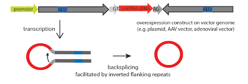
Overexpression of circRNA is not easy because of its low efficiency of loop forming, and it is easy to mismatch. By optimizing the binding sites of RBP, such as Alu elements and QKI, the circRNA can be formed accurately and efficiently. After overexpression, it is necessary to detect successful loop-forming and linear mRNA expression. In order to study the loop-forming efficiency of a new circRNA expression system, the mouse circrtn4 gene was selected to express in a variety of cell lines (including HeLa, N2a, HEK293). According to the RT-qPCR results in different cell lines, the efficiency of the new vector system pCircRNA-DMo-Rtn4 is much higher than that of the common vector system (pCircRNA-BE-Rtn4) in these cell lines.


CRISPR-U™ high-efficiency gene editing system

Ubigene focuses on genome editing, CRISPR-U™ is a gene-editing technology developed by Ubigene, which is more efficient than common CRISPR/Cas9 technologies in gene targeting. The services related to circRNA editing introduced in this paper can be provided by Ubigene, including knockout, interference, overexpression and circRNA expression testing.

How to construct a KO cell line | Ubigene


CRISPR/Cas9 technology enables the rapid generation of loss‐of‐function mutations in a targeted gene and changed the way of scientists approach their research. A single cell is a harbor of mutations that can be used to establish a new cell line, thereby creating a CRISPR‐induced knockout clone. These clonal cell lines serve as crucial tools for exploring protein function, analyzing the consequences of gene loss, and investigating the specificity of biological reagents. However, the successful derivation of knockout clones is technically challenging and in practice, it isn’t that simple. Obtaining high quality and reproducible editing results may need experience and optimization.


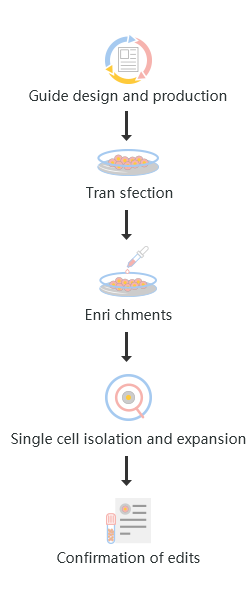
The standard workflow to generate CRISPR KO cell lines involves five major steps from project planning to confirmation of the desired edit. These steps are:

1. Design and production of KO guide

2. Cell transfection

3. Edited cells enrichments

4. Single-cell isolation and expansion

5. Confirmation of edits


Each of these steps requires careful consideration and technical skill in executing the many different methods involved.


KO gene editing workflow

Fig : KO gene editing workflow


Guide design and production:


The guide RNA recognizes the target gene region and directs the gene-editing machinery to the target section of DNA. For gRNA production, it is required to design and synthesize gRNA oligo sequence. The synthesized oligo was then cloned into a suitable expression vector and verified the oligo sequence.


A researcher can use a variety of guide formats to generate KO cell lines; single guide RNA (sgRNA), a two-part oligo system of cr:tracrRNA, in vitro-transcribed RNA, plasmid, and lentivirus. The plasmids and lentivirus- based approach are most popular to researchers, though they are time-consuming, require selection agents, and prone to off-target edits. However, lentivirus may offer advantages for difficult-to-transfect cells. The use of sgRNAs offers reduced off-target effects and time-saving benefits. Furthermore, the use of multiple gRNAs can increase the chance of obtaining the desired edit and save time downstream in clone selection stages. But these gRNAs must be targeted correctly. To begin with, gRNA targeting the gene of interest, the gRNA needs to clone in a suitable expression vector and sequence verification of gRNA.


Red Cotton™ CRISPR Gene Editing Designer is a free tool for knockout cell line strategy design. It contains parameters of 800 cell lines, and combine with Ubigene’s advanced gRNA design principle. Simply enter your target gene, and you could get 3 different KO strategies. There is a Red Cotton™ gRNA plasmid bank that has the validated gRNAs in stock. It contains over 10,000 gRNA plasmids for knockout cell line generation. Each plasmid only takes 80 USD.


Guide design and production:


When the CRISPR KO strategy has been finalized, choose the most efficient method for cell transfection is next. Determining the most efficient method is considered the most difficult step in the CRISPR workflow by many researchers. Each cell type often required an extensive optimization of the transfection conditions to accurately deliver the CRISPR gene-editing machinery into the cell. Besides that, some cell types, including stem cells, primary cells and immune and hematopoietic cell lineages are more difficult to transfect, as they have altered cell repair mechanisms, and lower cell viability. Thus, a successful transfection often depends upon previous experience with the cell line of choice to understand the cell characteristics.


Ubigene has worked on over 100 tyes of cell lines, and had developed a mature procedure to explore the optimal cell transfection method.


Enrichments and single cell expansion:


A clonal selection workflow is considered the best practice to develop a homogenous cell population that contains the desired KO. The researcher usually uses two common methods for enrichments and single cell expansion, one is dilution cloning and another, fluorescence-activated cell sorting (FACS) of single cells. Although both approaches can yield single cell–derived clones. FACS sorting is often more effective given its ability to specifically isolate double-positive cells. However, dilution cloning is cheaper and may cause less cellular stress than sorting. Furthermore, in this stage researcher should consider fully optimized growth conditions, otherwise, it could lead to the death of precious cell samples and the loss of all work in generating KO cell lines.


For some cell lines, generating sigle-cell clone is tricky, only a few clones may be able to thrive during isolation. Ubigene has rich experience on clonal isolation, so preliminary tests would be done to find out the best way to generate single-cell clones.


Confirmations of edits:


Once a KO cell line has been established, adequate validation of the specific gene edits is necessary. This requires the characterization of the KO and ensure the downstream viability of the work. Multiple methods offer the best assurance of an accurate gene edit. Common methods to validate engineered cell lines include Sanger sequencing, next-generation sequencing, and qPCR to verify the edit at a genomic level. Western blot and mass spectrometry can confirm of the KO at the proteomic level. For functional studies immunohistochemistry, immunocytochemistry and FACS are often used.


Ubigene CRISPR-U™ KO cell line:


Ubigene developed the CRISPR-UTM system for precise genome editing and our KO cell lines have been extensively validated using qPCR, Sanger sequencing, and in many cases, Western blotting to confirm full gene ablation at the genomic and proteomic levels. By using CRISPR-U™ developed KO cell line, researcher can reduce their experimental time and achieve target solutions quicker. Now Ubigene offer the best KO cell line service ever! Only 3480 USD, over 5000 genes guarantee WB result negative.


Reference:

Generating Single Cell–Derived Knockout Clones in Mammalian Cells with CRISPR/Cas9. Curr Protoc Mol Biol. 2019.

Tuesday, April 13, 2021

How to design gRNA and gRNA expression vector | Ubigene


The expression vector can drive high-level constitutive transcription of a user-selected gRNA sequence to achieve highly efficient CRISPR targeting when used in conjunction with Cas9 nuclease. A routine gRNA expression vector system would be simple and faster, cost-effective, efficient, and highly customizable. The development of a high-quality gRNA expression vector has been revolutionary and simplify CRISPR experiments.


1.What is reporter cell line and reporter gene?


Principle of gRNA design:


Reporters can be fluorescent and luminescent proteins such as green fluorescent protein (GFP) that exhibits bright green fluorescence when exposed to light in the blue to ultraviolet range, and the enzyme luciferase, which catalyzes a reaction with luciferin to produce light, in other cases, it can also be a tag that is to fused with the interested gene, such as glutathione S-transferase (GST), histidine (HIS), and flag tag, which will allow antibody-based detection and affinity-based isolation of the interested gene products.


CRISPR consists of an endonuclease protein (Cas9) for targeting a specific sequence RNA called guide RNA (gRNA) for guide endonuclease proteins to the target region. In CRISPR mediated genome editing, one of the key issue is to design and select the gRNA. Before starting to design and select gRNA, few questions need to address when choosing a gRNA: First, Does a given gRNA sequence exactly match your genomic target? If not the variation between a given gRNA sequence and genomic target may reduce the gRNA activity and increases the off-target effect. However, Cas9 enzyme can tolerate up to four mismatches (Between the gRNA and its genomic target), and one nucleotide gap. Thus, in a tolerable mismatches situation, cleavage can occur in the target site as well as a significant portion of off-target activity also occurs within gene coding sequences. Therefore, the off-target activity can lead to inaccurate interpretation of the CRISPR results and high off-target activity can cause depletion of the Cas9 enzyme or even cell death. Second, which species or variant of Cas9 (S. pyogenes, S. aureus, etc.) was this gRNA sequence designed for? A given gRNA sequence may only be compatible with a single species or PAM binding variant of Cas9. For instance, wild-type SpCas9 must be used with targets that are upstream of a 5' NGG 3' PAM sequence. Third, Which CRISPR application is this gRNA sequence compatible with? CRISPR knockout experiments use targeting sequences within exons, whereas CRISPR activation or repression experiments use targets within promoters.


There are multiple online tools are available to design guide RNAs with high editing efficiencies and low off-target effects. When designing gRNA by using online Bioinformatics tools, the following information is considered to choose the best gRNA. The gRNA with the lowest levels of Off-target, Optimum GC count, PAM (Protospacer Adjacent Motif), efficiency, different transcription variants of a gene, gRNA position, and no stable gRNA stem-loop. We’ve profiled the top CRISPR design tools that will simplify your experiments.


1. DESKGEN Cloud is the most comprehensive gRNA design software available, doing everything the MIT tool can and much more. By aggregating scoring functions from academic literature and other CRISPR tools, DESKGEN keeps you up to date with best practices in the field.


2. Benchling is designed with real scientist workflows in mind, from the most cutting-edge techniques to the most reliable everyday ones. Benchling product supports automated Gibson, Type IIS, and Digestion and Ligation cloning, so scientists do not need to switch between multiple tools.


3. MIT CRISPR design tool is a web tool crafted to simplify the process of CRISPR guide selection in an input DNA sequence by (i) discovering possible off-targets genome-wide, (ii) highlighting guides with high target specificity, and (iii) flagging guides with numerous or genic off-targets in target genomes.


4. CRISPR Gene Editing Designer (Red CottonTM) a computational tool to assist researchers in performing CRISPR-Cas9 experiment optimally. Red CottonTM provide maximize on-target activity (guide efficiency) as well as minimizing potential off-target effects (guide specificity) by analyzing the features of the target site. It is a very simple and user-friendly designing tools. The only species name, gene name/ID, and cell name are required to operate the system. The researcher can get valuable information about genes and cells within one minute with 3 knockout strategies, analysis of gene transcripts and sequence complexity, more than 800 cell line parameters, and 3000 successful knockout information.


Design gRNA with Red CottonTM tools


1. Design gRNAs using online tools Red CottonTM (https://www.rc-crispr.com/.).


2. Use the species name, gene name/ ID, and cell line name to generate potential gRNA sequences.


Note: All the potential gRNAs against the gene are created. The quality of the potential gRNAs will be reported as Score indicated on the on-target cutting activity.


3. Consider the Off-target activity in the gRNA selection when ranking the gRNAs. Note: Optimal guides are those with the greatest on-target efficiency and the least off-target activity. To disable a gene using Cas9 cutting, gRNAs are designed to target the first 10 - 50% of the coding sequence downstream of the initiating codon (as gRNAs targeting the 3’ end of the gene are less effective).


4. Ensure no internal BsmBI sites are present if oligonucleotides are being used for the Golden Gate cloning, as this will result in the annealed oligonucleotides being digested.


Using the Red CottonTM web tool (https://www.rc-crispr.com/), find candidate gRNA target sequences in the genomic region of interest. The output window shows 23 bp genomic sites of the form 5'-N20NGG-3' within your target region. These sites may reside on the + or - strand.


After select and synthesize gRNA oligos, it is necessary to construct a gRNA expression vector. The expression vector will be used for the transfection of cells.


gRNA expression vector (Plasmid):


The plasmid gRNA expression vector is a highly efficient tool for transfection-based delivery of target site-specific gRNA sequences into mammalian cells. Delivering plasmid vectors into mammalian cells by transfection is one of the most widely used procedures in bio-medical research. The plasmid transfection approach remains the workhorse of gene delivery due to its technical simplicity as well as good efficiency in a wide range of cell types. A key feature of transfection with plasmid vectors is that it is transient, with only a very low fraction of cells stably integrating the plasmid in the genome (typically less than 1%).


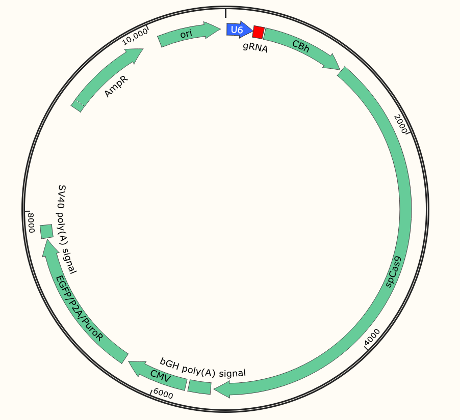
To achieve CRISPR-mediated gene targeting it is essential for the target cells to co-express both Cas9 as well as the target site-specific gRNA at the same time. One strategy of transfection is the vector carrying both Cas9 and the gRNA sequence and transfect the target cells, this transfection system is termed as an all-in-one vector transfection system. The vector carries reporter and selection markers (eg. EGFP and Puro).


 All in one plasmid vector

Fig 1: All in one plasmid vector


The other strategy is plasmid-based transfection by using separate vectors for driving Cas9 and gRNA expression (Cas9 only and gRNA only vectors respectively).


plasmid vector with single gRNA

Fig 2: plasmid vector with single gRNA


There are several advantages of using separate vector systems. Separate vectors offer the flexibility of combinatorial usage of different gRNA expression vectors in conjunction with a variety of Cas9 variants (wild type nuclease, nickase, nuclease-dead) depending upon the user’s experimental goal. Using a separate gRNA vector allows cells or organisms stably expressing high levels of Cas9 to be transfected with different gRNA sequences targeting either the same gene or different genes. This provides the opportunity for comparing the efficiencies of different gRNA sequences in parallel at CRISPR-mediated gene targeting in cells or organisms with comparable and high levels of Cas9 expression.

 Application:

1. The vector carrying reporter marker (eg. EGFP, Mcherry) can be used to monitor transfection efficiency and/or to enrich/isolate transfected cells by flow cytometry.


2. Selection markers (eg. Puro, Neo) confer resistance to antibiotics and helps for artificial selection.


Since the release of Red Cotton™ CRISPR Gene-editing Design System, it has brought the benefits to researchers from more than 20 countries and regions and assisted their gene study researches. In 2021, Ubigene continues to achieve our goal – “Make genome editing easier” and to bring more benefits to you.


gRNA plasmid bank

  Ubigene has set-up our gRNA plasmid bank!

  10000 in-stock plasmids available! Only $80.

  Deliver in one 3-5 week days.

  Most economical and lowest cost for gene KO.

  Check out your interested genes from our database now.

Click Here >
FREE plasmids

  gRNA plasmid bank plus Red Cotton system, the best and easiest kit for gene KO.

  Over 600 types of gene plasmids are now ready for you!

  Sign-up Red Cotton™ now and get your free vector. The first 50 registers can get the FREE plasmid each week.
Click the link to check out our free plasmids

Click Here >

Wednesday, April 7, 2021

Reporter KI cell model application | Ubigene


Since it was developed in 2012, this gene-editing tool has revolutionized biology research, making it easier to study disease and faster to discover drugs. The CRISPR/Cas9 system has become synonymous with genome editing in today’s world due to its simplistic mechanism. The CRISPR system is a precise genome editing technique, which can create gene knockout or knock-in genome manipulations through the substitution of a target genetic sequence with a desired donor sequence. Reporter KI is one of the most common applications of CRISPR/Cas9.


1.What is reporter cell line and reporter gene?


Reporter cell lines are stable cell lines that have been labeled with reporters, allowing the visualization, tracking, and isolation of interested genes. A reporter cell line can be generated through exogenous expression of a cassette with an easy-to-check tag (e.g. GFP) driven by the promoter of the interested gene. However, due to the exogenous expression level can lead to artifacts, a reporter with endogenous expression is preferred. In this regard, reporter cell line generated by knock-in a tag (e.g. GFP) downstream of the promoter of the interested gene is optimal.


Reporters can be fluorescent and luminescent proteins such as green fluorescent protein (GFP) that exhibits bright green fluorescence when exposed to light in the blue to ultraviolet range, and the enzyme luciferase, which catalyzes a reaction with luciferin to produce light, in other cases, it can also be a tag that is to fused with the interested gene, such as glutathione S-transferase (GST), histidine (HIS), and flag tag, which will allow antibody-based detection and affinity-based isolation of the interested gene products.


2.Strategy for reporter cell line generation by knock-in


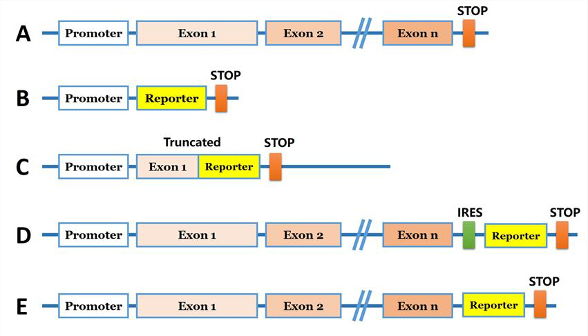
For the introduction of foreign DNA, the reporter gene are needed to be brought into the genome. Researchers can introduce a reporter coding sequence (e.g., eGFP) downstream of the promoter of the targeted gene (Fig 1B). Another strategy, the reporter coding sequence used to replace specific locus of the target gene and generate a truncated protein (Fig 1C).  A reporter gene can be inserted into a genomic site immediately downstream of the coding region of an endogenous gene, and co-expressed bi-cistronically with the endogenous gene as a single transcript under the control of the native transcriptional regulatory machinery (Fig. 1D). Furthermore, reporter gene coding sequence can be introduced in N/C terminal before the stop codon and under endogenous promoter control (Fig 1E). The strategies are commonly used for generation of a knock-in cellular models are shown in Fig 1.


Strategies for reporter gene knock-in

Fig 1: Strategies for reporter gene knock-in


3.Applications of reporter cell models


Transcriptional activity of gene promoter


Cell lines with endogenously tagged genes enable researchers to track protein production and localization in real time in live cells. These cell lines are also suitable for western blotting, protein pull down, affinity chromatography, immunocytochemistry, and flow cytometry among others. These can also be a good solution when working with a protein for which there is no good antibody. The targeted gene coding sequence is replaced with a reporter gene coding sequence (e.g., EGFP), where the endogenous gene is knocked out as the reporter gene is simultaneously knocked-in via homologous recombination. Upon recombination, the promoter of the targeted gene will drive expression of the inserted reporter gene. An example, luciferase knock-in to study transcriptional regulation of the human SREBP1 gene. In this study, the researcher integration of a single copy of the exogenous luciferase gene into one allele of the genome and a 14 bp deletion of the targeted sequence in the other. Luciferase activity was directly correlated with the promoter activity of the endogenous SREBP1 gene in the HEK293-SREBP1- T2A-luciferase-KI cell line cell line.


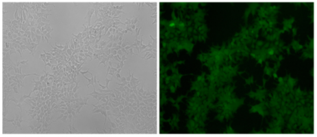
The HEK293 cell line expressed the exogenous eGFP reporter gene with undetectable expression of mCherry fluorescence after positive and negative selections with G418 and GCV.

Fig 2: The HEK293 cell line expressed the exogenous eGFP reporter gene with undetectable expression of mCherry fluorescence after positive and negative selections with G418 and GCV.


Expression, localization, and transportation of proteins in cells


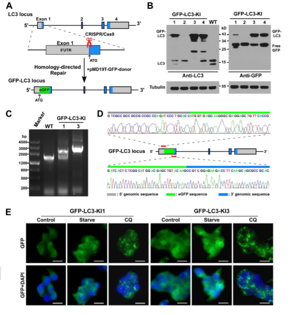
The visualization of a gene's expression can be realized by adding a tag sequence into the end (or the beginning) of the coding sequence of the targeted gene which will result in a fusion protein. As a result, expression levels of both the endogenous and recombinant proteins can be continuously monitored using fluorescence. The production of any protein of interest can be quantified based on the fluorescent reporter output of a cells. An example of knock-in, GFP-LC3 reporter in 293FT cells, where GFP is added to the N-terminal of endogenous LC3. Thus, the construction of the GFP-LC3 reporter knock in allowing GFP-LC3 fusion protein expression which is driven by the endogenous promoter without altering expression of the target gene. The endogenous GFP-LC3 reporter system showed accurate identification and quantification of cellular autophagy activity. The endogenous GFP-LC3 reporter system were verified by stress induced autophagy as well as drug (Rapamycin) induced/inhibit autophagy studies. As shown by the authors, free GFP protein is increase and its signal is still dispersed but stronger upon induction of autophagy, whereas GFP-LC3 become puncta like upon autophagy inhibition.


Generation of GFP-LC3 knock-in at the MAP1LC3B locus in 293FT cell.

Fig 3: Generation of GFP-LC3 knock-in at the MAP1LC3B locus in 293FT cell.


Target identification and evaluation of drug candidates


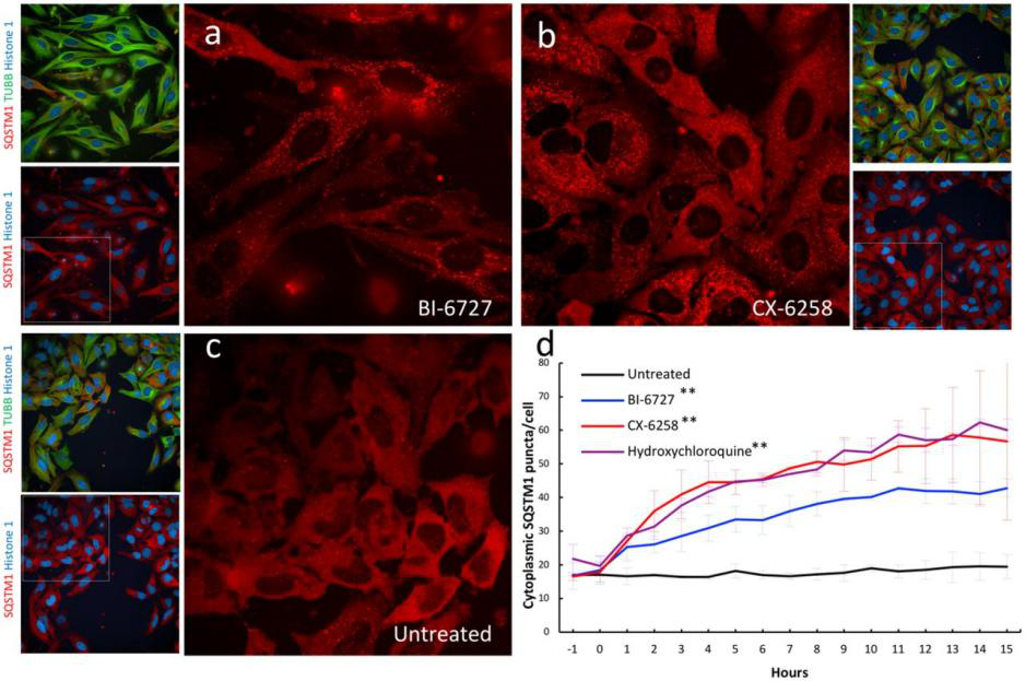
Genetically engineered live cells lines containing reporters for the expression of single specific genes is useful for drug target identification and candidate evaluation. Thus, once a particular gene has been chosen as a target, the first step of the process is to clone the regions of DNA that regulate expression of that gene and fused to a highly sensitive reporter gene like the firefly luciferase gene, which generates a readily measurable light signal in response to changes in transcription of the target gene. When an engineered cell line treated with appropriate compound generate that generate a light signal which reflects a specific change in gene expression. Therefore, a reporter cell-based assays can allow the rapid screening of a large number of samples and excluded those compounds that either cytotoxic or fail to interact with the cell. An example of reporter cell line drug target evaluation, a HELA cell line was tagged with a nuclear protein (Histone 1-mtagBFP2), a cytoskeleton marker (β tubulin-mClover3), and a known autophagy receptor protein (SQSTM1-mRuby3) to screen for kinase inhibitors (with known targets) that induce the accumulation of cytoplasmic autophagic vesicles for evaluating the accumulation of autophagic vesicles. The cell was treated with two kinase inhibitors PLK1 inhibitor BI-6727 and PIM kinases inhibitor CX-6258 that increase the number of autophagic vesicles like hydroxychloroquine, a known autophagy inhibitor (Fig 4).


Validation of kinase inhibitors that induce the accumulation of autophagic vesicles in a triple labeled HELA cell line.

Fig 4: Validation of kinase inhibitors that induce the accumulation of autophagic vesicles in a triple labeled HELA cell line.


In summary, reporter cellular models generated by knock-in are robust for functional studies such as monitoring gene expression and subcellular localization and are useful for high-through-put drug screening.

Ubigene has modified over 100 types of cell lines with our CRISPR-U system. It has optimized the gRNA designing and targeting constructs backbone. The efficiency of splicing and recombining would be much higher than the traditional CRISPR. Now we offer an affordable reporter KI cell line service, only 9980 USD, positive clone deliverable!

Tuesday, April 6, 2021

[Frontier Research] New method of spinal cord injury treatment | Ubigene

Mechanical injury of spinal cord tissue causes primary injury, and secondary neuromuscular response occurs in SCI after primary injury, mediating additional extensive nerve injury. Controlling harmful acute nerve injury can be used as a therapeutic strategy to inhibit injury and promote functional recovery. Microglia play a vital role in secondary injury after spinal cord injury. In the acute phase, activated microglia can produce neurotoxic pro-inflammatory cytokines, such as TNF-α, IL-1 cell killing factor and IL-6. therefore,.Carefully adjusting the functions of microglia to minimize their harmful effects and exert their neuroprotective effects is essential to promote the recovery of the nervous system.


Recently, researchers from Fujian Medical University published Inhibition of leucine rich repeats and calcium homology domain containing 1 accelerates microglia mediated neuroimaging in a rat traumatic spinal cord injury in The Journal of Neuroinflammation. In order to elucidate the significance of leucine-rich repeats and calponin homology domain containing 1 (LRCH1) on microglia function, researchers used lentivirus induced LRCH1 knockdown in primary microglia, and tested the role of LRCH1 in microglia mediated inflammation in vitro and in rat SCI model. It provides new clues for the study of novel treatments of SCI.


The expression of LRCH1 in microglia was down regulated after traumatic spinal cord injury. LRCH1 knockdown increases the generation of pro-inflammatory cytokines, such as IL-1 cytokines, TNF-α and IL-6. In addition, LRCH1 knockdown promoted microglia polarization to microglia with pro-inflammatory inducible nitric oxide synthase (iNOS) expression. LRCH1 knockdown also enhanced microglia mediated N27 neuronal death. Microglia infected by lentivirus carrying LRCH1 shRNA sequence is more toxic to primary rat spinal cord neurons ( purchased from Ubigene ).

Further study showed that LRCH1 knockdown increased the activation of p38 mitogen-activated protein kinase (MAPK) and Erk1/2 signaling, which are essential for the inflammatory response of microglia. When LRCH1 knockdown microglia were injected into the spinal cord of rats, they enhanced the production of pro-inflammatory cytokines, increased SCI induced leukocyte recruitment, aggravated SCI induced tissue injury and neuronal death, and worsened locomotor function.


In conclusion, studies have shown that inhibition of LRCH1 can increase the production of pro-inflammatory cytokines by activated microglia. In addition, inhibition of LRCH1 promoted the polarization of microglia to pro-inflammatory status. The effect of LRCH1 is mediating Erk1/2 signaling by p38 MAPK reduction. The low expression of LRCH1 and the adoptive transfer of microglia aggravate the tissue injury and dysfunction caused by spinal cord injury. Therefore, inhibition of LRCH1 can accelerate microglial neuroinflammation after spinal cord injury. For the first time, LRCH1 serves as a negative regulator of microglia-mediated neuroinflammation after SCI and provides clues for developing novel therapeutic approaches against SCI.


Ubigene provides LRCH1 knockout or knockdown services.

The editing efficiency of CRISPR-U™ system, exclusively developed by Ubigene, is 10 times higher than that of traditional methods. We have successfully performed gene knockout in over 100 types of cell lines, including various brain and nervous system cell lines, such as Human Neuroblastoma Cell Line SK-N-SH, Rat Glioblastoma Cell Line C6, Mouse Hippocampal Neuron Cell Line HT22, etc. Contact us now to learn more about your research related services!


Wednesday, March 31, 2021

Gene-Editing Salmonella: an ideal vector for tumor-targeting therapy | Ubigene

Salmonella is a facultative anaerobic gram-negative rod-shaped bacteria. Salmonella belongs to Enterobacteriaceae and is an important medical pathogen of humans and animals. Salmonella forms a complex bacterial community consisting of two species and six subspecies, including more than 2579 serotypes. At present, there are two species of Salmonella, S. entreica and S. bongori. The selective targeting of tumor tissue by Salmonella also makes it an ideal vector for tumorAs an intracellular parasite, Salmonella can effectively replicate and inhibit tumor growth in tumor tissue. After genetic engineering, it can be used as a carrier of tumor gene therapy in vitro and in vivo for liver cancer, gastric cancer and colorectal cancer.


 Salmonella and Tumor Therapy


1. Direct antitumor therapy with attenuated Salmonella


In view of the characteristics of Salmonella that can effectively inhibit tumor growth, scientists use a variety of genetic engineering techniques to modify the chromosome genome of Salmonella, which can reduce the virulence of Salmonella, so as to obtain the attenuated strain. While reducing the pathogenicity to the host, it still keeps high immunogenicity, thus ensuring the safety of clinical application.


2.Tumor targeting gene therapy


Attenuated Salmonella can carry exogenous genes, cytokines and exogenous effector proteins to treat tumors. Cytokines play an anti-tumor role by killing tumor cells directly. Exogenous effector proteins can be effectively transferred by attenuated Salmonella and expressed therapeutic proteins

 Knockout PhoP to construct attenuated Salmonella, improves the safety of   Salmonella in tumor treatment


The scientists used gene-splicing PCR combined with λ-Red system to delete PhoP from wild-type Salmonella typhimurium. phoP is a transcription regulator and a component of two regulatory systems that play key roles in adaptation to the homeostelium and in macrophage survival. It also controls the expression of more than 40 genes required by Salmonella typhimurium and its resistance to adverse environments in the host, such as low pH in the stomach, bile salt, hypoxia in the small intestine, and cationic antimicrobial peptides on epithelial cells. The disruption of phoP of Salmonella typhimurium leads to its inability to survive in phagocytes, and increases its sensitivity to stress, so as to achieve the function of reducing toxicity, and develop a safer bacterial therapy.


The phoP of wild type and standard strain of Salmonella typhimurium was disrupted by SOEing PCR. Three standard PCR and one fusion PCR were used to construct the linear DNA containing the upstream and downstream of phoP sequence and kanamycin cassette. pKD4 carrying kanamycin flanked by FRT (FLP recognition target) was used as a template plasmid. PCR confirmed that kanamycin had replaced phoP gene. Therefore, attenuated Salmonella was successfully constructed by removing phoP gene.


Gene-editing bacteria bring infinite possibilities to research, clinical medicine agriculture and other industrial productions!


Gene knockout has been used for many purposes, such as studying gene function, vaccine production and improving protein structure or expression. At present, the widely used traditional gene-editing methods include R6K suicide plasmid and λ-Red system.

Although the λ-Red system seems simple and has been successfully applied to E.coli and other gram-negative bacteria, the performance of this system in different bacteria is unstable due to the inherent differences of bacteria. In addition, suicide plasmid has some disadvantages such as host limitation and resistance residue. Other shortcomings of traditional KO include low recombination efficiency, heavy workload on validation, and residual loxP or FRT sites.


CRISPR/Cas9 technology is the most rapidly developed gene-editing technology in recent years. However, due to the lack of a repair system in bacteria, only a few microbial species were published to modify genes with this technology.Ubigene developed CRISPR-B™ technology, which is highly efficient in genome editing. CRISPR-B™ has features of easy-to-handle, accurate targeting, low off-target effect, scarless. It is efficient in various bacteria. The efficiency is more than 20x higher than that of traditional methods, easily achieve gene knockout, point mutation and knockin.

 A new treatment for tumor--CTNNB1-shRNA expression in Salmonella


Bacterial therapy has been used clinically for decades with a proven track record of safety in the treatment of gastroenteric diseases (for example, diarrhea, irritable bowel syndrome and inflammatory bowel disease). Moreover, recently there has been renewed interest in the clinical applications of live bacteria which is various non-pathogenic anaerobic bacteria are given intravenously and can infiltrate and replicate within solid tumors, particularly to treat human solid tumors. RNA interference (RNAi) has been established as an important research tool with great potential for gene therapy. Using a combination of bacterial therapy and RNAi therapy, scientists developed bacteria-mediated RNAi that delivers shRNA-expressing vectors to target cells to silence disease-causing genes.


Scientists have established a pSLS plasmid system that can express shRNAs in attenuated Salmonella. shRNA against HIV tat gene, human and mouse CTNNB1 genes were inserted into the multiple cloning site of pSLS. Then these plasmids were transformed into attenuated Salmonella, resulting in corresponding SL-pSLS-TAT, SL-pSLS-huCAT and SL-pSLS-mCAT strains. To determine whether SL-PSLS-HucAT can knockdown CTNNB1 expression of SW480 and inhibit the growth of SW480, SW480 cells were treated at different MOI.


CTNNB1 gene silencing significantly reduced cell proliferation and death in SW480 cells compared with control cells.To determine whether shRNA-expressing Salmonella could mediate anticancer effects in vivo, researchers performed studies using the SW480 xenograft tumor model. For these experiments, BALB/c female nude mice with established SW480 xenograft tumors were randomized into three groups to receive phosphate-buffered saline (PBS), SL-pSLS-TAT or SL-pSLS-huCAT. Tumor growth, as well as the expression levels of CTNNB1 and its downstream target genes c-Myc and cyclin D1, were recorded over a period of 2 weeks. Significantly, the growth of xenograft tumors treated with SL-pSLS-huCAT was reduced by 65% compared with tumors from the PBS control group, and by 45% compared with tumors treated with SL-pSLS-TAT . Reduced c-Myc and cyclin D1 protein levels were also observed in tumors treated with SL-pSLShuCAT compared with those treated with PBS. Thus,a significant reduction in tumor CTNNB1 protein level was observed when the mice were treated with SL-pSLS-huCAT.These results revealed that SL-pSLS-mCAT reduced the mRNA level of CTNNB1 in small intestines (by 34%), in polyps (by 73%) and in mucosal tissues (by 83%,) when compared with SL-pSLS-TAT-treated mice. The greater reductions observed in polyps and mucosal tissues over those found in the small intestines suggest a more selective invasion of these former tissues by Salmonella.


Taken together, these data suggest that attenuated shRNA-expressing Salmonella may be a powerful new tool for in vitro gene silencing, functional genomics, and the development of RNAi-based anticancer or human immunodeficiency virus therapeutics.


Ubigene developed CRISPR-B™ which optimizes the microbial gene-editing vectors and process. The efficiency and accuracy are 20x higher than traditional methods. CRISPR-B™ can be used in gene editing of bacteria and fungi. Contact us immediately to know about your research related services!


Reference:

1. A.Andino and I. Hanning. Review Article Salmonella enterica: Survival, Colonization, and Virulence Differences among Serovars. 2015. e Scientifific World Journal. 

2. Zhu Xiaozhou, Kong Guimei, Wan Dan, sun Guozhuang, Jiao Hongmei, Yin Yinyan, Li Guocai. Research progress of attenuated Salmonella in digestive system tumors. 2017. World Chinese Journal of digestion. 1480-1485

3. H Guo, J Zhang and C Ina. Targeting tumor gene by shRNA-expressing Salmonella-mediated RNAi. Gene Therapy. 2011. 18:95-105 

4. Ahani Azari, A. Zahraei Salehi, T. Nayeri FasaeiB.and Alebouyeh, M..Gene disruption in Salmonella typhimurim by modified λ Red disruption system. Iranian Journal of Veterinary Research ,Shiraz University. 2015. 52:301-305


[Research highlight] Enhancing p53 pathway can efficiently suppress colon cancer

  Colorectal cancer is the third most diagnosed cancer and leads to the second mortality among cancers worldwide. The first-line chemotherap...